
Pancreatic ductal adenocarcinoma (PDAC) is not only genetically complex; it is metabolically adaptive. Under nutrient limitation, oxidative stress, and tissue-level microenvironmental pressure, pancreatic cancer cells can rewire lipid metabolism to maintain membrane structure, redox balance, signaling, and survival. Lipidomics in pancreatic cancer research provides a direct way to measure these lipid remodeling events and connect them with stress adaptation, drug response, and target discovery. This article explains how lipidomics can reveal targetable metabolic vulnerabilities, using a recent Nature Communications study on cystine limitation stress in PDAC as a featured example, and discusses how bulk, spatial, and multi-omics approaches can support cancer metabolism research.
1. Why Lipid Metabolism Matters in Pancreatic Cancer
PDAC develops within a biologically demanding environment. Dense stromal architecture, poor vascularization, hypoxia, nutrient limitation, and inflammatory signaling can all influence how pancreatic cancer cells access and use metabolic resources. In this context, lipid metabolism is more than a background process. It affects membrane biogenesis, energy storage, signaling, stress tolerance, and cell-state transitions.
Cancer cells may alter fatty acid synthesis, phospholipid remodeling, sphingolipid metabolism, lipid droplet dynamics, cholesterol handling, and redox-linked lipid pathways. These changes can help cells adapt to stress, but they may also create dependencies. When a tumor cell becomes unusually reliant on a specific metabolic route, that route can become a research target or a candidate vulnerability for further mechanistic investigation.
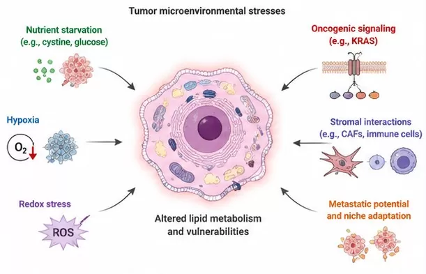
Figure 1. Multiple stress conditions can reshape cancer cell lipid metabolism and expose metabolic vulnerabilities. Adapted from Park et al., The Heterogeneity of Lipid Metabolism in Cancer, in The Heterogeneity of Cancer Metabolism, Springer, 2021. Licensed under CC BY 4.0.
This is why lipidomics has become increasingly important in cancer metabolism research. Instead of measuring a single lipid or pathway marker, lipidomics profiles many lipid species across classes, allowing researchers to see coordinated patterns of remodeling. These patterns can reveal how cancer cells adapt, which pathways are perturbed, and which lipid changes are associated with treatment response or stress sensitivity.
2. Cystine Limitation Stress: A Window into Cancer Cell Vulnerability
Cystine availability is closely linked to cysteine metabolism, glutathione synthesis, and antioxidant defense. Because glutathione helps cells manage oxidative stress, cystine limitation can pressure cancer cells at the intersection of nutrient availability, redox homeostasis, and lipid metabolism. When antioxidant capacity is disrupted, lipid peroxidation and lipid remodeling can become especially relevant to cell survival.
Stress conditions are useful in cancer metabolism studies because they may reveal dependencies that are not obvious under standard culture conditions. A cancer cell that appears metabolically flexible under nutrient-rich conditions may become vulnerable when a specific nutrient, antioxidant pathway, or lipid remodeling route is limited. Lipidomics helps researchers observe the downstream molecular consequences of that stress.
In pancreatic cancer, this type of stress-based analysis is particularly relevant because PDAC cells often operate in a metabolically constrained microenvironment. By comparing lipid profiles across stress and control conditions, researchers can identify lipid class shifts, pathway-level remodeling, and candidate metabolic dependencies that may merit deeper study.
3. Featured Study: Lipidomics Identifies a Targetable Vulnerability in PDAC
A recent Nature Communications study that cited targeted quantitative lipidomics support from MetwareBio investigated how adaptation to cystine limitation stress can create a targetable lipid metabolism vulnerability in pancreatic ductal adenocarcinoma. The study focused on pancreatic cancer cells and used lipidomics to characterize lipid metabolism changes associated with stress adaptation.
The significance of this study is not simply that lipid levels changed. The broader lesson is that lipidomics can help researchers detect how cancer cells reorganize lipid metabolism under selective pressure. In the context of cystine limitation, lipid profiles can provide evidence of altered membrane lipid composition, stress-associated remodeling, and lipid pathways linked to cellular survival strategies.
For drug discovery and translational research, this type of evidence is valuable because it moves the analysis from phenotype to mechanism. If a stress condition exposes a lipid metabolism dependency, researchers can investigate whether that dependency is reproducible, pathway-specific, and biologically meaningful. In this way, lipidomics can support target discovery, drug response studies, biomarker research, and multi-omics mechanism exploration.
Featured publication: Adaptation to cystine limitation stress confers a targetable lipid metabolism vulnerability in pancreatic ductal adenocarcinoma. Nature Communications. Read the publication on PMC.
4. What Lipidomics Can Reveal in Cancer Metabolism Studies
Lipidomics is most informative when lipid changes are interpreted as biological patterns rather than isolated molecular lists. In cancer metabolism studies, several layers of interpretation are especially useful.
4.1 Lipid Class-Level Changes
Different lipid classes can point to different biological processes. Phospholipids reflect membrane composition and remodeling. Sphingolipids can be involved in stress signaling, inflammation, and cell fate regulation. Glycerolipids can indicate lipid storage and energy adaptation. Sterol lipids are related to membrane organization and signaling, while fatty acyls may connect to oxidation, inflammatory mediators, and lipid peroxidation biology.
4.2 Pathway-Level Lipid Remodeling
The strongest lipidomics studies usually go beyond differential lipid screening. A shift in phosphatidylcholine, phosphatidylethanolamine, ceramide, triglyceride, or cholesterol ester patterns becomes more useful when it is connected to pathway activity, enzyme regulation, transport processes, redox status, or treatment effects. This is where lipidomics begins to support mechanism-driven cancer research.
4.3 Drug Response and Metabolic Vulnerability Signals
In drug response studies, lipidomics can compare treated and untreated samples to determine whether a perturbation affects lipid metabolism. In vulnerability-focused studies, lipidomics can help identify lipid features associated with stress sensitivity, resistance, or adaptation. These lipid patterns do not automatically become biomarkers or drug targets, but they can provide strong candidates for follow-up validation.
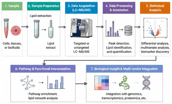
Figure 2. Mass spectrometry-based lipidomics workflow from sample preparation to data acquisition, annotation, statistical analysis, and biological interpretation. Adapted from Züllig, Trötzmüller, and Köfeler, Lipidomics from sample preparation to data analysis: a primer, Analytical and Bioanalytical Chemistry, 2020. Licensed under CC BY 4.0.
Table 1. Lipid classes and their relevance in cancer metabolism
| Lipid Class | Biological Relevance | Potential Cancer Research Insight |
|---|---|---|
| Phospholipids | Membrane structure, signaling, remodeling | Stress- or treatment-associated membrane adaptation |
| Sphingolipids | Stress signaling, inflammation, cell fate | Cell-state changes and vulnerability-associated pathways |
| Glycerolipids | Energy storage and lipid droplets | Nutrient stress adaptation and lipid storage dynamics |
| Sterol lipids | Membrane organization and cholesterol metabolism | Growth signaling and lipid homeostasis |
| Fatty acyls | Oxidation, inflammatory mediators, lipid peroxidation context | Redox-linked lipid remodeling and stress response |
Table 2. What lipidomics can reveal in cancer metabolism studies
| Research Question | What Lipidomics Can Reveal |
|---|---|
| How do cancer cells adapt to nutrient stress? | Lipid class shifts, membrane remodeling, and stress-associated lipid changes |
| Is a treatment affecting lipid metabolism? | Treatment-associated lipid changes and pathway-level remodeling |
| Are lipid changes linked to redox or cystine stress? | Lipid remodeling patterns related to oxidative stress and lipid homeostasis |
| Are molecular changes localized in tissue? | Spatial lipid distribution when combined with spatial lipidomics |
| Which mechanisms may explain lipid changes? | Connections between lipids, genes, proteins, and pathways through multi-omics integration |
5. From Bulk Lipidomics to Spatial Lipidomics in Cancer Research
Bulk lipidomics measures lipid abundance from homogenized samples. It is powerful for comparing groups, treatments, or stress conditions, but it averages signals across all cells and regions within a sample. For many cancer studies, that average can hide important spatial biology.
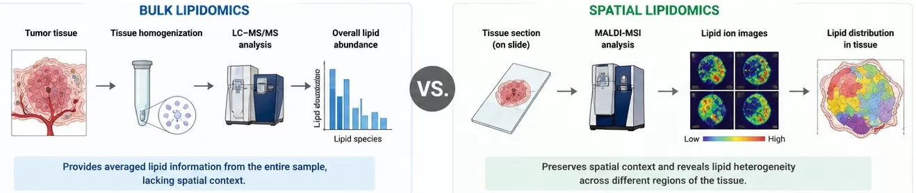
Figure 3. Bulk lipidomics vs. spatial lipidomics in cancer tissue.
Tumors are heterogeneous. Lipid remodeling may differ between tumor regions, stromal areas, necrotic zones, invasive fronts, immune-rich regions, or treatment-affected tissue. Spatial lipidomics adds tissue localization by mapping lipid distributions directly within tissue sections using mass spectrometry imaging workflows. This can help researchers ask not only which lipids changed, but where those changes occurred.
For pancreatic cancer and other solid tumor models, spatial lipidomics can complement bulk lipidomics when tissue architecture matters. It may help investigate tumor microenvironment-associated lipid patterns, region-specific lipid remodeling, drug response effects, and spatially resolved biomarker candidates. When combined with spatial metabolomics, spatial proteomics, or transcriptomics, it can contribute to a more tissue-aware view of cancer biology.
6. FAQ on Lipidomics in Pancreatic Cancer Research
6.1 Why Is Lipidomics Useful in Pancreatic Cancer Research?
Lipidomics is useful because pancreatic cancer cells often rewire lipid metabolism to support membrane remodeling, stress adaptation, signaling, and survival. Measuring lipid changes helps researchers identify altered lipid pathways, treatment-associated remodeling, and potential metabolic vulnerabilities for follow-up mechanistic studies.
6.2 What Is a Targetable Metabolic Vulnerability?
A targetable metabolic vulnerability is a pathway or dependency that cancer cells rely on under specific conditions. If that dependency becomes important for survival, proliferation, or stress adaptation, it may become a candidate for target discovery, drug response studies, or mechanism-focused research.
6.3 How Is Cystine Limitation Connected to Lipid Metabolism?
Cystine availability is connected to cysteine metabolism, glutathione synthesis, and antioxidant defense. When cystine is limited, cells may experience redox stress and altered lipid homeostasis, which can lead to measurable lipid remodeling. Lipidomics provides a way to capture these downstream changes.
6.4 Is Lipidomics the Same as Metabolomics?
Lipidomics is a specialized branch of metabolomics focused on lipids. General metabolomics captures a broad range of small molecules, while lipidomics uses lipid-focused extraction, separation, annotation, and analysis strategies to better characterize lipid classes such as phospholipids, sphingolipids, glycerolipids, and sterol lipids.
6.5 When Should Researchers Consider Spatial Lipidomics?
Researchers should consider spatial lipidomics when tissue context is central to the question. In cancer research, this includes tumor microenvironment studies, region-specific lipid remodeling, localized drug response, or spatial biomarker discovery. Spatial lipidomics complements bulk lipidomics by showing where lipid changes occur within tissue sections.
6.6 Can Lipidomics Be Integrated with Proteomics or Transcriptomics?
Yes. Integrating lipidomics with proteomics or transcriptomics can help connect lipid changes with upstream enzymes, transporters, signaling pathways, and regulatory mechanisms. This is especially useful in cancer metabolism research, where lipid remodeling may reflect broader changes in gene expression, protein abundance, and pathway activity.
How MetwareBio Supports Lipidomics and Cancer Metabolism Research
MetwareBio supports lipidomics, metabolomics, proteomics, spatial omics, and multi-omics analysis for life science research. For cancer metabolism studies, lipidomics can help characterize lipid remodeling under stress, treatment, disease-model, or genetic perturbation conditions. Spatial lipidomics can further add tissue localization when the study involves tumor architecture, microenvironmental heterogeneity, or region-specific molecular changes.
If you are interested in lipidomics or cancer metabolism research, please do not hesitate to contact us.
Contact UsRead More: Lipidomics, Cancer Metabolism & Multi-Omics
Explore these related articles to deepen your understanding of lipidomics workflows, lipid biology, spatial omics, and cancer metabolism research — from foundational concepts to advanced multi-omics integration strategies.
New to lipidomics? This guide walks you through the core concepts — from lipid classification and extraction strategies to LC-MS/MS workflows and data interpretation. It provides an accessible entry point for researchers looking to incorporate lipidomics into cancer or disease metabolism studies.
After acquiring lipidomics data, the analytical challenge begins. This step-by-step guide covers quality control, normalization, differential lipid screening, lipid class-level pattern analysis, and pathway enrichment — exactly the workflow needed to translate raw lipidomics output into biological insight.
Understanding which lipid classes are relevant to your research question is critical for designing and interpreting lipidomics experiments. This article covers 13 major lipid classes including phospholipids, sphingolipids, glycerolipids, and sterol lipids, detailing their structures, biological functions, and analytical approaches.
Spatial metabolomics and spatial lipidomics share the same tissue-imaging principle. This article explains how mass spectrometry imaging maps metabolite distributions within tissue sections, discusses its role in tumor microenvironment studies, and provides context for designing spatially resolved cancer metabolism experiments.
Multi-omics integration of proteomics and lipidomics is increasingly used in metabolic disease and cancer research. This article explores how combining protein-level and lipid-level data can reveal enzyme-metabolite relationships, dysregulated pathways, and mechanistic connections that neither platform captures alone.
Fatty acid metabolism is reprogrammed across multiple cancer types, not only in pancreatic cancer. This article examines how lung cancer cells exploit lipid metabolism changes for survival, proliferation, and treatment resistance — providing a comparative perspective on lipid vulnerability research in solid tumors.
References
- Li Y, Li Z, Li Q, et al. Adaptation to cystine limitation stress confers a targetable lipid metabolism vulnerability in pancreatic ductal adenocarcinoma. Nature Communications. 2026;17:1343. https://doi.org/10.1038/s41467-025-68099-0
- Hanahan D. Hallmarks of Cancer: New Dimensions. Cancer Discovery. 2022;12(1):31-46. https://doi.org/10.1158/2159-8290.CD-21-1059
- Pavlova NN, Thompson CB. The Emerging Hallmarks of Cancer Metabolism. Cell Metabolism. 2016;23(1):27-47. https://doi.org/10.1016/j.cmet.2015.12.006
- Snaebjornsson MT, Janaki-Raman S, Schulze A. The multifaceted role of lipid metabolism in cancer. Cell Metabolism. 2020;31(1):62-76. https://doi.org/10.1016/j.cmet.2019.11.010
- Beloribi-Djefaflia S, Vasseur S, Guillaumond F. Lipid metabolic reprogramming in cancer cells. Oncogenesis. 2016;5:e189. https://doi.org/10.1038/oncsis.2015.49
- Dixon SJ, Lemberg KM, Lamprecht MR, et al. Ferroptosis: an iron-dependent form of nonapoptotic cell death. Cell. 2012;149(5):1060-1072. https://doi.org/10.1016/j.cell.2012.03.042
- Viswanathan VS, Ryan MJ, Dhruv HD, et al. Dependency of a therapy-resistant state of cancer cells on a lipid peroxidase pathway. Nature. 2017;547(7664):453-457. https://doi.org/10.1038/nature23007
- Jiang X, Stockwell BR, Conrad M. Ferroptosis: mechanisms, biology and role in disease. Nature Reviews Molecular Cell Biology. 2021;22:266-282. https://doi.org/10.1038/s41580-020-00324-8
- Ellis SR, Paine MRL, Eijkel GB, et al. Automated, parallel mass spectrometry imaging and structural identification of lipids. Nature Methods. 2018;15:515-518. https://doi.org/10.1038/s41592-018-0010-6
