Skin and mucosal surfaces encounter bacteria, fungi, viruses, and environmental agents every day, yet most exposures do not become invasive infections. Antimicrobial peptides (AMPs) are part of this first-line defense. Some are present at baseline, whereas others are released after epithelial stress or microbial sensing. Defensins are one important AMP family, alongside cathelicidins, dermcidin, histatins, and related host-defense peptides. Calling them “natural antibiotics” captures their ability to inhibit microbes but misses much of their biology. AMPs may disrupt membranes, trap pathogens, recruit immune cells, alter cytokine signaling, shape microbial communities, and support tissue repair. Their activity also changes with the surrounding matrix, including salt concentration, pH, mucus, proteins, and proteases. This article examines the main human AMP families, their membrane and non-membrane mechanisms, their roles at epithelial barriers, disease-associated dysregulation, and the contribution of peptidomics and multi-omics to AMP research (Mookherjee et al., 2020; Oliveira Júnior et al., 2025).
1. Defensins and Antimicrobial Peptides: Core Families and Biological Features
1.1 Antimicrobial Peptides as Innate Host-Defense Effectors
Most well-studied human AMPs are short and positively charged, with hydrophilic and hydrophobic regions that favor membrane interactions. This pattern is common rather than universal. AMP length, net charge, folding, disulfide content, and post-translational processing vary considerably, as do their cellular sources. Epithelial cells, neutrophils, macrophages, Paneth cells, sweat glands, and salivary glands each contribute distinct peptide pools.
Many AMPs are not released in their final active form. They are translated as precursors and activated by proteolytic cleavage, so transcript abundance alone cannot show which mature peptides are present or active. The term “host-defense peptide” is often used when immune signaling, barrier maintenance, and tissue repair are central to the biology (Mookherjee et al., 2020; Hellinger et al., 2023).
LL-37 illustrates precursor-to-mature AMP processing.
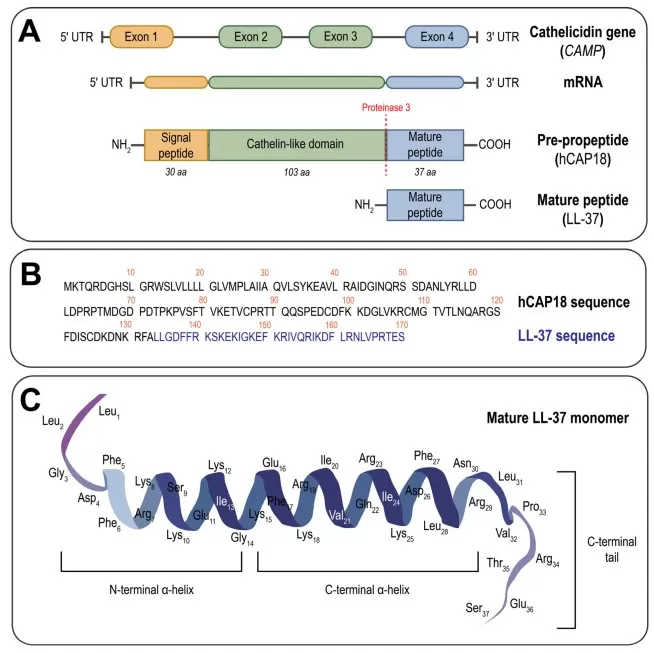
Figure 1. CAMP/hCAP18 organization and proteolytic release of mature LL-37. Reproduced from Voronko et al. (2025), International Journal of Molecular Sciences, 26(16), 8103, under CC BY 4.0; resized without content changes.
1.2 Major Human Defensins and Other Antimicrobial Peptide Families
Human defensins are cysteine-rich peptides whose folds are stabilized by disulfide bonds. Alpha-defensins include neutrophil HNP1-4 and intestinal HD5/HD6; beta-defensins are produced mainly at epithelial surfaces. LL-37, dermcidin-derived peptides, and salivary histatins extend the human AMP repertoire beyond defensins and differ in tissue source and dominant function (Mookherjee et al., 2020; Johnstone & Herzberg, 2022).
Table 1. Major Human AMP Families, Sources, and Functions
| Peptide family | Representative peptides | Major sources | Research relevance |
|---|---|---|---|
| Alpha-defensins | HNP1-4 | Neutrophils | Phagocyte-associated antimicrobial defense |
| Alpha-defensins | HD5, HD6 | Intestinal Paneth cells | Crypt protection and mucosal defense |
| Beta-defensins | hBD-1 to hBD-4 | Epithelial cells | Skin and mucosal immunity |
| Cathelicidins | LL-37 | Epithelial and immune cells | Antimicrobial and immunomodulatory activity |
| Dermcidin | DCD-derived peptides | Sweat glands | Skin-surface defense |
| Histatins | Histatin family | Salivary glands | Oral and antifungal defense |
2. How Antimicrobial Peptides Work: Microbial Selectivity, Membrane Disruption, and Non-Lytic Functions
Membrane disruption is the best-known AMP mechanism, but no single model fits every peptide. Activity shifts with peptide concentration and aggregation, microbial species and membrane composition, pH, ionic strength, and surrounding proteins or extracellular matrix.
2.1 Electrostatic Selectivity for Microbial Membranes
Negatively charged phospholipids, lipopolysaccharide, lipoteichoic acids, and wall teichoic acids make many microbial surfaces favorable targets for cationic AMPs. After electrostatic attraction brings a peptide to the surface, amphipathic regions can associate with the lipid-water interface and sometimes enter the bilayer. Fungi and enveloped viruses present different lipid and surface chemistries, which helps explain why activity spectra vary among peptides.
Mammalian membranes differ from microbial membranes in outer-leaflet lipids, cholesterol, and surface charge, creating relative rather than absolute selectivity. Greater hydrophobicity or high peptide concentrations may increase cytotoxicity and hemolysis. Salts, serum proteins, mucus, extracellular DNA, and local pH can also reduce the amount of free peptide available for membrane binding (Zhang et al., 2021; Oliveira Júnior et al., 2025).
2.2 Carpet, Barrel-Stave, and Toroidal-Pore Models
Three models are commonly used to describe membrane permeabilization. Surface-bound peptides destabilize the bilayer in the carpet model; barrel-stave assemblies form peptide-lined pores; and toroidal pores are lined by both peptides and curved lipid headgroups. Real membranes often show transient defects or mixed states, so these models are useful descriptions rather than strict categories (Zhang et al., 2021; Oliveira Júnior et al., 2025).
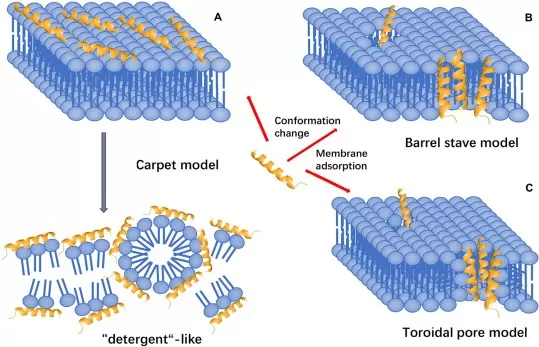
Figure 2. Carpet, barrel-stave, and toroidal-pore models of AMP-mediated membrane disruption. Reproduced from Huan et al. (2020), Frontiers in Microbiology, 11, 582779, under CC BY 4.0; resized without content changes.
2.3 Beyond Membrane Lysis: Intracellular Targets, Microbial Trapping, and Immune Modulation
Some AMPs enter or perturb microbial membranes without causing immediate lysis. Once inside, they may interfere with cell-wall synthesis, ribosomes, nucleic acids, protein folding, or enzymes; others reduce adhesion or biofilm formation. Human alpha-defensin 6 follows a different strategy: it self-assembles into nanonet-like structures that trap bacteria in the intestinal lumen (Chu et al., 2012).
Direct killing is only part of the response. AMPs can recruit leukocytes, neutralize endotoxin, alter cytokine signaling, promote epithelial migration, and support wound repair. A peptide may therefore matter in vivo even when it shows modest killing in a simplified assay. Studies should report whether the observed endpoint is killing, growth inhibition, trapping, antibiofilm activity, or host-cell signaling rather than treating all of them as the same “antimicrobial” effect (Mookherjee et al., 2020).
3. Defensins and Antimicrobial Peptides in Epithelial Barrier Immunity
At healthy barriers, AMPs work alongside epithelial junctions, mucus, ciliary or fluid clearance, resident immune cells, and commensal organisms. Together, these defenses keep microbes away from vulnerable tissue without sterilizing the surface.
3.1 Skin Barrier Defense and Wound Repair
Keratinocytes, resident immune cells, hair follicles, and sweat glands produce overlapping sets of AMPs. Human beta-defensins, LL-37, and dermcidin-derived peptides help limit colonization in the acidic, lipid-rich environment of the skin. Some also influence keratinocyte migration, immune-cell recruitment, angiogenesis, and wound closure. Their effects vary with anatomical site, injury, sweat and sebum composition, and the resident microbiota (Mookherjee et al., 2020).
3.2 Intestinal Crypt Defense and Microbiota Compartmentalization
Paneth cells at the base of small-intestinal crypts release HD5, HD6, lysozyme, and other effectors close to the stem-cell niche. This concentrated secretion limits microbial access to regions of epithelial renewal. Alpha-defensins also help organize host-microbiota spatial relationships, supporting compartmentalization rather than indiscriminate clearance (Wallaeys et al., 2023). HD6 adds a physical layer of defense by forming nanonets that trap microbes (Chu et al., 2012).
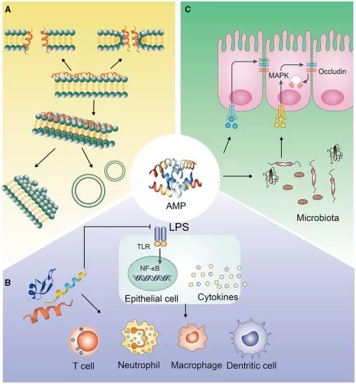
Figure 3. Generalized schematic of direct antimicrobial, immune-regulatory, and epithelial-protective AMP pathways at the intestinal barrier. Reproduced from Gong et al. (2021), Frontiers in Nutrition, 8, 751010, under CC BY 4.0; resized without content changes.
3.3 Coordinated AMP Defense Across Respiratory, Oral, and Urogenital Mucosa
Respiratory epithelia secrete beta-defensins and LL-37 into a system that also relies on mucus, mucociliary clearance, surfactant-associated factors, and recruited leukocytes. In the oral cavity, histatins and defensins work with salivary flow and mucosal integrity, with activity against both bacteria and fungi. Urogenital epithelia likewise deploy defensins and cathelicidin as part of locally regulated mucosal defense. At each site, peptide activity is shaped by secretion, diffusion, proteolysis, binding partners, and physical clearance (Johnstone & Herzberg, 2022).
4. Antimicrobial Peptide Dysregulation in Infection and Inflammatory Disease
An AMP increase is not automatically protective, and a decrease does not always indicate weaker defense. Pathogens may evade peptide attack, the local matrix may inactivate the peptide, and host production may be too low, excessive, or misplaced. These processes can reinforce barrier damage, dysbiosis, and persistent inflammation.
4.1 Pathogen Evasion and Microenvironmental Inactivation of AMPs
Bacteria evade AMPs by changing surface charge or membrane lipids, producing capsules, increasing efflux, releasing proteases, or sequestering peptides in extracellular material. Biofilms add diffusion and binding barriers and can shift cells into more tolerant physiological states. Heritable resistance, reversible tolerance, and loss of activity caused by a salt-, protein-, or protease-rich matrix are biologically distinct and should be reported separately (Tajer et al., 2024).
In chronic wounds and persistent airway infections, detecting LL-37 or a defensin does not show how much intact peptide reaches the pathogen. Proteolysis, oxidation, matrix binding, ionic inhibition, and spatial separation can all lower effective exposure (Mookherjee et al., 2020; Tajer et al., 2024).
4.2 AMP Dysregulation: Deficiency, Excess, and Mislocalization
Low production or weak induction can compromise local control, but persistently high expression can also intensify chemotaxis, inflammatory signaling, or tissue injury. Processing, secretion, degradation, and tissue localization further separate total peptide abundance from effective activity.
Skin disease provides clear examples. Atopic dermatitis combines barrier dysfunction, type 2 inflammation, dysbiosis, and altered AMP responses that can favor Staphylococcus aureus colonization (Peng et al., 2026). Psoriasis often shows strong AMP induction in already inflamed tissue. In rosacea and chronic wounds, LL-37 processing, protease activity, or peptide persistence may affect both inflammation and microbial defense (Mookherjee et al., 2020).
4.3 Barrier Damage, Dysbiosis, and Interpreting AMP Changes
Barrier injury increases microbial access and exposes AMPs to altered pH, salts, proteases, plasma proteins, and extracellular DNA. If the peptide layer is weakened or compositionally altered, microbial encroachment can sustain epithelial stress and inflammation. Similar feedback is seen in Paneth-cell dysfunction, chronic wounds, and recurrent airway infections (Mookherjee et al., 2020; Johnstone & Herzberg, 2022; Wallaeys et al., 2023).
Interpreting a measured change requires its compartment and disease context. Tissue abundance, secreted concentration, and circulating levels are not interchangeable, and interpretation should consider the producing cell, anatomical site, disease stage, treatment, microbiome, pathogen burden, and barrier integrity. Higher abundance may reflect effective defense, compensation, or inflammatory amplification; lower abundance may arise from reduced production, loss of producing cells, degradation, or dilution. Mechanistic interpretation requires both identification of the mature peptide and evidence of its biological activity.
5. Therapeutic Development of Antimicrobial Peptides: Opportunities, Barriers, and Engineering
AMP-derived therapeutics are being developed both to kill microbes and to modify host responses such as inflammation, immune-cell recruitment, and repair. Most practical formats emphasize local exposure: topical agents, wound dressings, antimicrobial coatings, delivery systems, antibiotic adjuvants, and peptidomimetics. Local delivery avoids some of the demands of prolonged systemic circulation, and combination use may improve membrane penetration, weaken biofilms, or reduce the dose of a conventional antimicrobial (Mookherjee et al., 2020; Cesaro et al., 2023; Oliveira Júnior et al., 2025).
Development is difficult because peptides can be rapidly degraded, cleared, or bound by serum proteins; activity may fall in physiological salt, and cytotoxicity, hemolysis, manufacturing, delivery, and resistance also require attention. Sequence optimization, cyclization, D-amino-acid substitution, lipidation, peptidomimetics, nanoparticles, hydrogels, and functionalized surfaces are being used to improve stability, selectivity, and local exposure (Cesaro et al., 2023; Oliveira Júnior et al., 2025). For now, AMP-derived molecules are better viewed as complementary tools for topical, biomaterial-associated, host-directed, or combination therapy than as universal antibiotic replacements. Each candidate still needs pharmacology, toxicity, resistance, and efficacy testing in the intended biological setting.
6. Peptidomics and Multi-Omics for Defensin and Antimicrobial Peptide Research
AMP abundance is shaped by transcription, precursor synthesis, proteolytic maturation, secretion, chemical modification, tissue distribution, and degradation. Biological activity depends on the local tissue and microbial environment, while measured activity also varies with assay conditions. Peptidomics measures endogenous peptide forms directly; other omics layers help identify their cellular source, regulation, and biological setting.
6.1 Why Mature-Peptide Measurement Requires Peptidomics
RNA-seq and qPCR measure DEFA, DEFB, CAMP, and related transcripts, but they do not reveal which mature peptides are present. One precursor may yield several cleavage products, and oxidation, truncation, disulfide formation, and other modifications can change stability or activity. Conventional bottom-up proteomics digests proteins with trypsin, whereas endogenous peptidomics aims to recover peptides already present in the sample (Hellinger et al., 2023).
LC-MS/MS peptidomics can identify sequences and cleavage sites, compare peptide abundance, and reveal forms not predicted from gene-level data. Recovery and identification remain difficult for low-abundance peptides, closely related isoforms, highly cationic sequences, and disulfide-rich defensins. A non-detection result is therefore not proof of biological absence.
6.2 Pre-Analytical and Analytical Considerations for AMP Peptidomics
Potential research matrices include skin or intestinal tissue, wound fluid, saliva, sputum or bronchoalveolar lavage fluid, culture supernatant, and organoid-conditioned media. Because endogenous peptide recovery is highly matrix-dependent, sample feasibility, stabilization strategy, extraction conditions, and expected peptide abundance should be confirmed during study design. Collection and processing must suppress ex vivo proteolysis and artificial modification. Rapid cooling or stabilization, validated protease control, low-binding consumables, standardized extraction, limited freeze-thaw cycles, randomized processing, pooled quality controls, and batch monitoring are key design elements (Hellinger et al., 2023).
Record pH, salt concentration, hemolysis or cellular contamination, extraction recovery, and whether disulfide bonds were preserved, reduced, or alkylated. Peptide abundance does not measure antimicrobial activity. Functional assays should specify the microorganism and growth state, medium, salt and protein content, peptide concentration, exposure time, and endpoint so that results can be interpreted alongside LC-MS/MS data.
Figure 4 shows a published wound-fluid workflow that combines peptide extraction, LC-MS/MS, peptide identification, protease analysis, AMP prediction, and data visualization (Hartman et al., 2021).
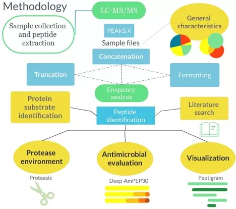
Figure 4. Representative wound-fluid peptidomics workflow combining peptide extraction, LC-MS/MS, peptide identification, protease analysis, AMP prediction, and visualization. Reproduced from Hartman et al. (2021), Frontiers in Immunology, 11, 620707, under CC BY 4.0; resized without content changes.
6.3 Integrating Peptidomics with Complementary Omics Layers
Transcriptomics measures the expression of defensin genes, CAMP, cytokines, epithelial differentiation programs, and proteases. Proteomics adds information on barrier proteins, immune effectors, processing enzymes, and tissue-damage markers, while peptidomics identifies mature AMP forms and cleavage products. Microbiome profiling describes community composition and pathogen burden, and metabolomics characterizes microbial metabolites, nutrient availability, and redox-associated metabolites in the local environment.
Table 2. Sample and Multi-Omics Strategies for AMP Research
| Research question | Suggested sample | Recommended strategy | Main interpretation |
|---|---|---|---|
| Skin barrier and wound infection | Skin tissue or wound fluid for host peptide/protein profiling; swabs for microbiome analysis | Peptidomics/proteomics on host samples + matched swab microbiome profiling | AMP processing, colonization, and tissue damage |
| Intestinal defensin biology | Intestinal tissue or organoids for host omics; stool or mucosal samples for microbiome analysis | Transcriptomics + peptidomics on host samples + matched microbiome profiling | Paneth-cell activity and host-microbiome interactions |
| Airway infection | Sputum or BALF for secreted peptides and microbes; epithelial models for mechanism studies | Peptidomics + inflammatory protein or transcriptomic readouts + transcriptomics or microbiome analysis as appropriate | Mucosal AMP response and inflammation |
| Mechanism validation | Cells, organoids, and defined synthetic-peptide systems | Peptide analysis + activity assays + proteomics | Direct antimicrobial and host-cell effects |
Each omics layer should test a specific part of the proposed mechanism. Reduced HD5 abundance, for example, can be examined together with Paneth-cell markers, protease expression, microbiome encroachment, and intestinal phenotype. In wound fluid, LL-37 forms can be compared with biofilm signatures, host proteases, inflammatory proteins, and healing outcomes. These comparisons can distinguish changes in peptide production from altered processing, degradation, microbial escape, or downstream tissue responses.
7. Frequently Asked Questions About Defensins and Antimicrobial Peptides
Are defensins the same as antimicrobial peptides?
No. Defensins are one major AMP family, alongside cathelicidins, dermcidin-derived peptides, and histatins. They are defined by conserved cysteines and disulfide-stabilized folds, whereas other AMP families differ in structure, source, and function (Mookherjee et al., 2020).
How do antimicrobial peptides kill or control microbes?
Many cationic AMPs bind negatively charged microbial surfaces and disrupt membranes. Others act on intracellular targets, inhibit biofilms, trap microbes, or modulate host immunity. Which mechanism dominates depends on the peptide, microorganism, concentration, and local environment (Zhang et al., 2021; Oliveira Júnior et al., 2025).
Can antimicrobial peptides replace conventional antibiotics?
Not broadly. AMP-derived therapies may suit topical delivery, wound care, coatings, or combination treatment, but stability, toxicity, delivery, manufacturing, and resistance remain important barriers. They are better viewed as complements to conventional antibiotics than as universal replacements (Cesaro et al., 2023; Oliveira Júnior et al., 2025).
Why is peptidomics useful for defensin and AMP research?
Gene and conventional protein measurements may not reveal which mature endogenous peptides are present. Peptidomics can identify cleavage products and compare peptide forms and abundance, but functional assays are still needed because abundance alone does not establish antimicrobial activity (Hellinger et al., 2023).
What samples are suitable for antimicrobial peptide peptidomics?
Potential samples include tissue, wound fluid, saliva, sputum or BALF, culture supernatant, and organoid-conditioned media, depending on the study question. Because endogenous peptides are sensitive to proteolysis and sample handling, matrix feasibility, stabilization strategy, and extraction workflow should be discussed before study launch.
Can RNA-seq replace peptidomics for defensin research?
No. RNA-seq can measure defensin genes, CAMP, cytokines, proteases, and epithelial-response programs, but it cannot determine which mature peptide forms are present. Peptidomics is useful because many AMPs are produced as precursors and become active only after proteolytic processing.
From AMP Biology to Study Design: How MetwareBio Supports Research
Study design should match the analytical strategy to the peptide form, sample matrix, and biological question. Endogenous peptide profiling can be combined with proteomics, transcriptomics, microbiome analysis, metabolomics, and multi-omics interpretation to connect mature peptide forms with upstream regulation and downstream biological context. When direct antimicrobial activity is central to the research question, peptidomics results should be interpreted together with separately designed functional validation assays.
MetwareBio offers 4D label-free LC-MS/MS peptidomics with ddaPASEF acquisition for endogenous peptide identification and relative quantification, together with complementary proteomics and multi-omics services.
Contact UsRead More: Peptidomics, Proteomics, and Multi-Omics for Peptide Research
These articles cover complementary topics for researchers studying defensins and antimicrobial peptides, from peptide sequencing methods and proteomics quality control to multi-omics integration in clinical research.
Explore the mass spectrometry methods used for peptide identification and sequencing, including how LC-MS/MS approaches support endogenous peptide discovery in peptidomics studies of defensins and other AMPs.
Learn how combining quantitative proteomics with PTM profiling reveals regulatory mechanisms in epithelial barrier signaling, immune cell activation, and AMP expression beyond protein abundance alone.
Compare serum and plasma sample types relevant to circulating AMP and defensin studies, including collection considerations for peptide stability and protease activity in blood-derived matrices.
Discover how metaproteomics characterizes microbiome-host interactions at mucosal barriers, complementing AMP peptidomics with microbial protein profiles in intestinal and skin microbiome studies.
Understand how QC samples, protease controls, and batch monitoring ensure reliable peptide identification and quantification in peptidomics studies of wound fluid, saliva, and other complex biological matrices.
See how multi-omics integration connects protein-level findings with metabolite profiles in clinical studies, providing a framework for combining AMP peptidomics with complementary omics in disease research.
References
- Cesaro, A., Lin, S., Pardi, N., & de la Fuente-Núñez, C. (2023). Advanced delivery systems for peptide antibiotics. Advanced Drug Delivery Reviews, 196, 114733. https://doi.org/10.1016/j.addr.2023.114733
- Chu, H., Pazgier, M., Jung, G., Nuccio, S. P., Castillo, P. A., de Jong, M. F., Winter, M. G., Winter, S. E., Wehkamp, J., Shen, B., Salzman, N. H., Underwood, M. A., Tsolis, R. M., Young, G. M., Lu, W., Lehrer, R. I., Bäumler, A. J., & Bevins, C. L. (2012). Human alpha-defensin 6 promotes mucosal innate immunity through self-assembled peptide nanonets. Science, 337(6093), 477-481. https://doi.org/10.1126/science.1218831
- Gong, T., Fu, J., Shi, L., Chen, X., & Zong, X. (2021). Antimicrobial peptides in gut health: A review. Frontiers in Nutrition, 8, 751010. https://doi.org/10.3389/fnut.2021.751010
- Hartman, E., Wallblom, K., van der Plas, M. J. A., Petrlova, J., Cai, J., Saleh, K., Kjellström, S., & Schmidtchen, A. (2021). Bioinformatic analysis of the wound peptidome reveals potential biomarkers and antimicrobial peptides. Frontiers in Immunology, 11, 620707. https://doi.org/10.3389/fimmu.2020.620707
- Hellinger, R., Sigurdsson, A., Wu, W., Romanova, E. V., Li, L., Sweedler, J. V., Süssmuth, R. D., & Gruber, C. W. (2023). Peptidomics. Nature Reviews Methods Primers, 3, 25. https://doi.org/10.1038/s43586-023-00205-2
- Huan, Y., Kong, Q., Mou, H., & Yi, H. (2020). Antimicrobial peptides: Classification, design, application and research progress in multiple fields. Frontiers in Microbiology, 11, 582779. https://doi.org/10.3389/fmicb.2020.582779
- Johnstone, K. F., & Herzberg, M. C. (2022). Antimicrobial peptides: Defending the mucosal epithelial barrier. Frontiers in Oral Health, 3, 958480. https://doi.org/10.3389/froh.2022.958480
- Mookherjee, N., Anderson, M. A., Haagsman, H. P., & Davidson, D. J. (2020). Antimicrobial host defence peptides: Functions and clinical potential. Nature Reviews Drug Discovery, 19(5), 311-332. https://doi.org/10.1038/s41573-019-0058-8
- Oliveira Júnior, N. G., Souza, C. M., Buccini, D. F., Cardoso, M. H., & Franco, O. L. (2025). Antimicrobial peptides: Structure, functions and translational applications. Nature Reviews Microbiology, 23, 687-700. https://doi.org/10.1038/s41579-025-01200-y
- Peng, G., Abudouwanali, A., Sun, Q., Tan, Y., Zhao, W., Yang, M., Wang, S., Ogawa, H., Okumura, K., & Niyonsaba, F. (2026). Role of antimicrobial peptides in the pathogenesis of atopic dermatitis. The Journal of Dermatology, 53(3), 372-379. https://doi.org/10.1111/1346-8138.17975
- Tajer, L., Paillart, J.-C., Dib, H., Sabatier, J.-M., Fajloun, Z., & Abi Khattar, Z. (2024). Molecular mechanisms of bacterial resistance to antimicrobial peptides in the modern era: An updated review. Microorganisms, 12(7), 1259. https://doi.org/10.3390/microorganisms12071259
- Voronko, O. E., Khotina, V. A., Kashirskikh, D. A., Lee, A. A., & Gasanov, V. A. O. (2025). Antimicrobial peptides of the cathelicidin family: Focus on LL-37 and its modifications. International Journal of Molecular Sciences, 26(16), 8103. https://doi.org/10.3390/ijms26168103
- Wallaeys, C., Garcia-Gonzalez, N., & Libert, C. (2023). Paneth cells as the cornerstones of intestinal and organismal health: A primer. EMBO Molecular Medicine, 15(2), e16427. https://doi.org/10.15252/emmm.202216427
- Zhang, Q.-Y., Yan, Z.-B., Meng, Y.-M., Hong, X.-Y., Shao, G., Ma, J.-J., Cheng, X.-R., Liu, J., Kang, J., & Fu, C.-Y. (2021). Antimicrobial peptides: Mechanism of action, activity and clinical potential. Military Medical Research, 8, 48. https://doi.org/10.1186/s40779-021-00343-2