Membrane transport proteins regulate the selective movement of ions, nutrients, metabolites, signaling molecules, and drugs across cellular and organelle membranes. Their activity shapes nutrient uptake, metabolic exchange, ion homeostasis, cell signaling, and the absorption, distribution, and efflux of many drugs. Because they sit at the interface between cellular physiology and pharmacology, transporters can function both as therapeutic targets and as determinants of efficacy, toxicity, and resistance.
However, transporter function cannot be inferred from gene or protein abundance alone. Membrane localization, substrate specificity, transport activity, downstream metabolic effects, and compound engagement represent distinct evidence layers that must be evaluated together. This article summarizes the major SLC and ABC transporter groups, together with representative pumps and channels; explains the core principles of substrate recognition, alternating access, and energy coupling; examines their roles in drug discovery, disposition, and resistance; and discusses how proteomics, metabolomics, isotope tracing, and functional assays can support mechanism-oriented research and drug-target validation.
1. Membrane Transport Proteins: Core Functions and Major Families
Membrane transport proteins are predominantly integral membrane proteins or membrane-embedded protein complexes that enable controlled molecular movement across the plasma membrane and organelle membranes. In this article, the term includes channels, carrier-type transporters, and pumps. In the sections below, transporter refers mainly to solute carrier (SLC), ATP-binding cassette (ABC), and other substrate-binding systems that move solutes through conformational cycles.
1.1 Selective Transport, Transport Modes, and Evidence Layers
Ions, sugars, amino acids, organic acids, nucleosides, lipid precursors, and many xenobiotics require dedicated transport routes. These routes create concentration differences between the cytosol, extracellular space, mitochondria, lysosomes, endoplasmic reticulum, and other compartments. The resulting gradients support nutrient acquisition, pH control, electrical excitability, neurotransmitter recycling, metabolic exchange, and waste elimination.
Transport biology must be interpreted at several levels. mRNA abundance reports steady-state transcript levels; protein abundance indicates how much transporter is detected; surface or organelle localization indicates where it may operate; and transport assays measure substrate movement. Metabolite profiling and isotope tracing provide complementary evidence of downstream biochemical consequences. These readouts are related, but not interchangeable.
Channels form aqueous pathways that allow selected ions or water to move rapidly, usually down an electrochemical gradient. Carrier-type transporters bind a substrate and alternately expose the binding site to opposite sides of the membrane. They may mediate facilitated diffusion, symport, or antiport. Pumps use a primary energy source, commonly ATP hydrolysis, to drive transport against a gradient. This mechanistic distinction is more informative than treating every membrane transport protein as the same type of molecular gate.
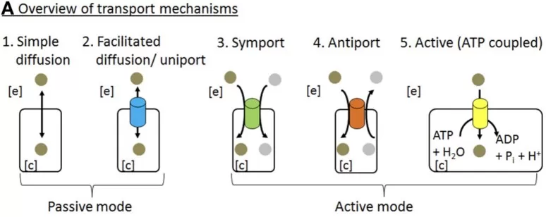
Figure 1. Major modes of molecular transport across the plasma membrane. The diagram summarizes simple diffusion, facilitated diffusion, symport, antiport, and ATP-coupled active transport. Cropped from Figure 1A in Sahoo et al. (2014), Frontiers in Physiology, under the Creative Commons Attribution License (CC BY). The image was cropped to retain panel A; no other content changes were made.
1.2 SLC Transporters: Nutrient, Metabolite, and Drug Transport
The SLC group contains many evolutionarily distinct families rather than one uniform structural class. Its members transport glucose, amino acids, monocarboxylates, nucleosides, bile acids, metals, neurotransmitters, and numerous drugs. Some work as uniporters, whereas others couple substrate movement to Na+, H+, or another solute gradient. Representative systems include GLUT1/SLC2A1, SGLT2/SLC5A2, LAT1/SLC7A5, xCT/SLC7A11, MCT1/SLC16A1, and the serotonin transporter SERT/SLC6A4. Their folds, oligomeric states, tissue distributions, and kinetic properties differ substantially (Pizzagalli et al., 2021).
This diversity creates both opportunity and complexity. SLC proteins can control nutrient dependency, metabolic exchange, neuronal signaling, and tissue exposure to drugs. However, close homologs may share substrates, and one transporter may recognize several related molecules. Functional annotation therefore requires more than a family name or sequence match.
1.3 ABC Transporters and Other Major Transport Systems
Transporting ABC proteins generally combine transmembrane domains with cytosolic nucleotide-binding domains. ATP binding and hydrolysis are coupled to conformational changes that move substrates across the membrane. ABC family members transport lipids, sterols, bile components, peptides, metabolites, and xenobiotics. ABCB1, ABCC1, and ABCG2 are especially prominent in drug efflux and multidrug-resistance research, but the wider superfamily includes proteins with different substrates, tissue distributions, structural organizations, and physiological roles (Sajid et al., 2023).
Primary pumps such as Na+/K+-ATPase, Ca2+-ATPases, H+/K+-ATPase, and V-type ATPases maintain ion gradients, organelle acidity, and membrane potential. Aquaporins and voltage- or ligand-gated ion channels provide rapid pathways for water or ions and are established pharmacological targets. They complete the broader transport landscape, while the mechanistic and omics sections below focus mainly on carrier-type SLC and ABC transporters.
Table 1. SLC vs. ABC Transporters: Energy Coupling, Substrates, Structure, and Drug Relevance
| Feature | SLC Transporters | ABC Transporters |
|---|---|---|
| Main energy coupling | No direct energy input for facilitated diffusion; pre-existing ion or solute gradients for secondary active transport | ATP binding and hydrolysis |
| Typical mechanisms | Uniport, symport, antiport; selected members show atypical channel-like or enzymatic functions | Primarily ATP-driven transport; functions vary across subfamilies |
| Representative members | GLUT1/SLC2A1; SGLT2/SLC5A2; LAT1/SLC7A5; xCT/SLC7A11; MCT1/SLC16A1; SERT/SLC6A4 | ABCB1 (P-gp); ABCC1 (MRP1); ABCG2 (BCRP) |
| Drug relevance | Nutrient dependence, drug uptake, metabolic exchange, neurological signaling | Drug efflux, multidrug resistance, xenobiotic and lipid transport |
| Research challenges | Substrate overlap among homologs; limited selective inhibitors; annotation gaps | Polyspecificity; species differences; conformational complexity |
2. Membrane Transporter Mechanisms: Structure, Alternating Access, and Energy Coupling
A transporter structure identifies the arrangement of helices, cavities, gates, and coupling elements, but transport is a dynamic process. Mechanistic interpretation requires connecting structural states with substrate binding, energy input, membrane conditions, and measurable movement across the bilayer.
2.1 Substrate Recognition and Structural Insights
Most human SLC and ABC transporters use multiple transmembrane alpha-helices to form a substrate-binding cavity and a controlled translocation pathway. Hydrogen bonds, electrostatic interactions, hydrophobic contacts, hydration, and local steric constraints shape substrate recognition. Binding pockets can be sufficiently flexible to accommodate related endogenous compounds or drugs, which helps explain substrate overlap but complicates selective inhibitor design.
Cryogenic electron microscopy has transformed transporter structural biology by resolving large membrane-protein complexes and capturing ligand-bound or nucleotide-dependent conformations that were difficult to obtain by crystallography alone. Structural series can reveal gates, coupling ions, lipid contacts, and candidate conformational intermediates along a proposed transport cycle. Nevertheless, a cryo-EM structure provides a conformational snapshot rather than a complete description of transport. Mutagenesis, transport kinetics, binding measurements, and complementary biophysical methods are needed to determine whether an observed state belongs to the active transport cycle (Baril et al., 2023).
2.2 Alternating Access, Energy Coupling, and Membrane Regulation
Carrier-type transporters commonly follow an alternating-access principle: the substrate-binding site is exposed to one side of the membrane, becomes occluded, and then opens to the opposite side. Outward-facing, substrate-bound, occluded, inward-facing, release, and reset states provide a useful general sequence. Rocker-switch, rocking-bundle, and elevator models describe different large-scale movements that can implement this principle. They should not be treated as a single universal mechanism for every transporter, channel, or pump.
ABC transporters also alternate access, but their transmembrane rearrangements are coupled to nucleotide binding and hydrolysis at the nucleotide-binding domains. The shared concept is controlled access to opposite membrane sides; the structural motion and source of energy are family specific.
Secondary active transporters use pre-existing Na+, H+, or solute gradients, sometimes together with membrane potential, to move another substrate uphill. Stoichiometry matters because the number and direction of coupled ions determine thermodynamic feasibility and transport direction. ABC transporters and primary pumps instead couple conformational cycling directly to ATP-dependent reactions.
The surrounding membrane is not an inert scaffold. Bilayer thickness, curvature, charge, and specific lipid interactions can stabilize particular conformations or alter kinetics. Oligomerization and accessory subunits may further change trafficking, substrate preference, or transport efficiency. Structural and biochemical studies increasingly show that ion and lipid coordination is integral to secondary active transport rather than a secondary detail (Drew & Boudker, 2024).
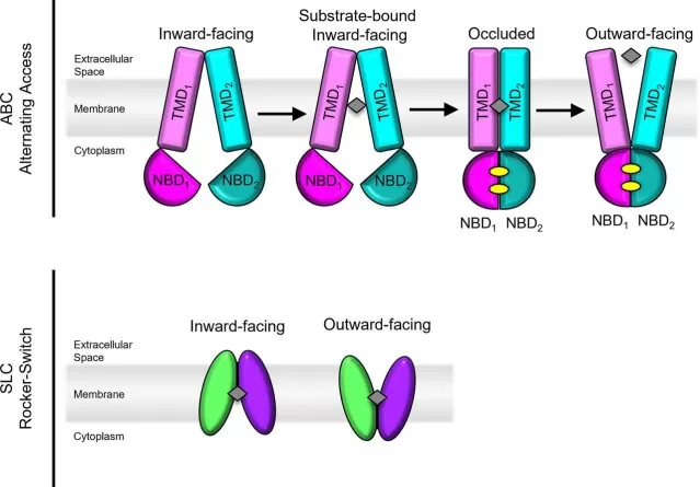
Figure 2. Representative alternating-access mechanisms in pharmacologically relevant ABC and SLC transporters. The upper panel illustrates ATP-coupled conformational cycling in ABCB1, ABCC1, and ABCG2, whereas the lower panel shows the rocker-switch mechanism described for SLC19A1 and SLC29A1. Reproduced from Baril et al. (2023), Drug Metabolism and Disposition, under the Creative Commons Attribution 4.0 International License (CC BY 4.0); resized for layout. No content changes were made.
3. Membrane Transporters in Drug Discovery and Resistance
Transport proteins influence therapeutics in two distinct ways. Some are intentionally modulated as direct targets. Others primarily determine how a drug enters cells, crosses tissue barriers, is eliminated, or is exported from resistant cells. Separating these roles prevents pharmacokinetic effects from being mistaken for direct target pharmacology.
3.1 Direct Therapeutic Targets, Drug Disposition, and Resistance
Several transporter classes demonstrate direct druggability. SGLT2 inhibition reduces renal glucose reabsorption, while monoamine transporter modulation changes neurotransmitter reuptake. H+/K+-ATPase inhibition suppresses gastric acid secretion. In cancer metabolism, LAT1/SLC7A5 and xCT/SLC7A11 are investigated because they can support amino acid acquisition and redox homeostasis, although context-dependent expression and metabolic redundancy complicate translation. Urate and bile-acid transporters provide additional examples in which manipulating solute handling may alter systemic metabolite balance (Galetin et al., 2024; Pizzagalli et al., 2021).
Uptake and efflux transporters in the intestine, liver, kidney, blood-brain barrier, and other tissues help determine drug exposure. Genetic variants, disease states, inflammation, diet, and co-administered compounds can change abundance or activity, creating variability in efficacy, toxicity, and drug-drug interactions. ABCB1, ABCC1, and ABCG2 can export structurally diverse agents and contribute to reduced intracellular drug accumulation in experimental resistance models (Galetin et al., 2024; Sajid et al., 2023).
These effects are difficult to predict from substrate lists alone. Transporters can overlap in tissue distribution and specificity, and the net disposition of a drug may reflect competing uptake, efflux, metabolism, and passive permeability. Experimental systems must therefore be matched to the question, and tissue exposure should not be inferred from plasma concentration alone.
3.2 Evaluating Membrane Transporters as Drug Targets
A practical evaluation asks whether the transporter is mechanistically necessary, pharmacologically accessible, and sufficiently selective. Key criteria include genetic or intervention-based evidence linking transport function to the phenotype; a defined substrate, transport direction, and measurable activity; expression in the relevant tissue or disease state; a tractable substrate pocket, allosteric site, or conformational state; limited compensation by homologous transporters; and an acceptable safety window when normal homeostasis or tissue barriers are perturbed. These criteria distinguish a disease-associated transporter from a viable therapeutic target. Binding and target engagement establish compound interaction, but direct transport modulation, selectivity, and phenotype rescue remain separate requirements.
4. Membrane Transporter Proteomics, Metabolomics, and Functional Validation
No single analytical layer fully describes a membrane transporter. Proteomics can identify abundance, localization, modifications, or partners; metabolomics can show substrate and pathway consequences; and functional assays can measure direct movement. The strongest designs use these readouts to answer different questions rather than treating cross-omics correlation as proof of mechanism.
4.1 Quantitative and Surface Proteomics for Abundance and Localization
Membrane proteins are analytically challenging because they are hydrophobic, often low in abundance, and unevenly recovered during extraction, digestion, and peptide detection. Global quantitative proteomics can screen for transporter changes across conditions, while targeted mass spectrometry can verify selected proteins with greater analytical focus. Calibrated assays may support absolute abundance estimates, but only when suitable standards and validated workflows are used (Achour et al., 2020).
Total abundance does not establish functional placement. Surface enrichment, cell-surface labeling, flow cytometry, imaging, or organelle-resolved proteomics can test whether a transporter reaches the relevant membrane. Post-translational modification (PTM) proteomics and interaction methods can investigate phosphorylation, ubiquitination, glycosylation, internalization, accessory proteins, and trafficking machinery. When compound-induced changes are under investigation, these measurements can be paired with orthogonal binding or target-engagement assays.
4.2 Metabolomics and Isotope Tracing for Metabolite Pools and Flux
Targeted metabolomics is useful when the candidate substrates and downstream products are known, whereas untargeted metabolomics can reveal broader pathway remodeling. A change in an intracellular or extracellular metabolite pool may support a transport hypothesis, but it can also result from altered synthesis, degradation, sequestration, cell composition, or parallel transport routes. Annotation confidence, matrix effects, normalization, and quality control therefore remain central to interpretation (Alseekh et al., 2021).
Stable-isotope tracing adds dynamic information by following labeled atoms from a supplied substrate into downstream metabolites. It can test substrate contribution, pathway branching, and metabolic rewiring that steady-state abundance cannot resolve. Even then, tracer selection, time course, compartmentation, exchange reactions, and modeling assumptions determine what can be concluded. Isotope labeling can support flux-oriented interpretation, but it does not replace a direct uptake, efflux, vesicle-transport, or ion-flux assay when the central claim concerns transport rate (Jang et al., 2018).
4.3 Functional and Causal Validation of Transport Mechanisms
Direct assays should measure the substrate, direction, rate, kinetics, and dependence on ions, ATP, membrane potential, or competing ligands. Cell-based uptake and efflux assays, inside-out or membrane-vesicle systems, electrophysiology, ion-sensitive readouts, and reconstituted proteoliposomes address different transporter classes. Appropriate negative controls, inactive analogs, expression controls, and orthogonal detection methods reduce the risk of attributing nonspecific toxicity or membrane damage to transporter inhibition. These functional measurements also provide the context needed to test whether structurally observed states participate in an active transport cycle (Baril et al., 2023).
Causality is strengthened when genetic perturbation changes the transport phenotype and re-expression restores it. Mutating a predicted binding or coupling residue can connect a structural model to function. Concordant proteomics, metabolomics, and phenotypic changes can then define the biological consequence. This layered approach also exposes disagreement: increased total protein with unchanged surface localization, for example, would suggest examining altered trafficking, retention, or inactive protein accumulation rather than assuming increased transport.
Table 2. Membrane Transporter Validation Methods: Evidence Layers, Readouts, and Limitations
| Research question | Recommended method | Main readout | Main limitation |
|---|---|---|---|
| Where and how strongly is the transporter expressed? | Quantitative proteomics | Relative abundance; absolute abundance with calibrated targeted assays | Low-abundance hydrophobic proteins may be under-detected; resolution depends on sampling |
| Is it accessible at the relevant membrane? | Surface proteomics, flow cytometry, imaging, or organelle proteomics | Localization and accessibility | Enrichment, antibody, accessibility, and compartment-purity bias |
| What are the substrate and transport direction? | Uptake/efflux assays, vesicle transport, electrophysiology | Substrate, rate, direction, kinetics | Cell context, assay format, and detection sensitivity vary |
| Does transport change the metabolic phenotype? | Metabolomics and isotope tracing | Metabolite pools and flux | Indirect; alternative routes and compartmentation |
| Is the transporter a viable drug target? | Compound screening + transport assay + selectivity testing | Engagement, inhibition, selectivity | Off-target effects, resistance, and in vivo translation |
| Does genetic evidence support causality? | Knockdown/KO + rescue + mutagenesis | Phenotype linkage and structure-function | Redundancy, compensation, and model limitations |
| What is the structural basis of transport? | Cryo-EM, crystallography, cross-linking, modeling | Conformational states and binding sites | Static snapshots; dynamic cycle requires functional validation |
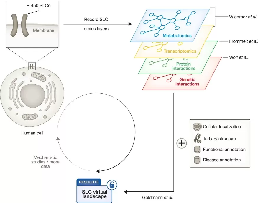
Figure 3. Integrating multi-omics layers to characterize solute carrier biology. Metabolomics, transcriptomics, protein-interaction, and genetic-interaction datasets are integrated with cellular localization, structural, functional, and disease annotations to build a systematic reference landscape for human SLC biology. Reproduced from Billmann (2025), Molecular Systems Biology, under the Creative Commons Attribution 4.0 International License (CC BY 4.0); resized for layout. No content changes were made.
5. Integrated Workflow for Membrane Transporter Research
A disciplined workflow begins with the biological question rather than a preferred platform. Candidate discovery may combine disease genetics, transcriptomics, quantitative proteomics, metabolomics, literature evidence, and structural information. Protein abundance and localization should then be verified in the relevant sample, cell type, and membrane compartment before direct transport assays establish substrate movement, direction, kinetics, and energy dependence. Metabolomics and isotope tracing can subsequently define the biochemical consequences of the transport phenotype.
Structural and omics evidence become most informative when linked through testable hypotheses. Structural approaches, including cryo-EM, can nominate candidate substrate-contact, gating, or coupling residues. Quantitative and surface proteomics can establish abundance and localization, while site-directed mutagenesis and direct transport assays test structural predictions. Metabolomics or isotope tracing then connects transport changes to downstream pathway consequences.
How MetwareBio Supports Membrane Transporter and Multi-Omics Research
Membrane transporter projects often require a staged strategy that distinguishes candidate discovery from functional proof. MetwareBio can support protein-level screening through quantitative proteomics, focused verification with PTM proteomics. Integrated <span ;"="">transcriptomics, proteomics, and metabolomics can assess candidate substrate pools, coordinated regulation, compensatory responses, and phenotype-linked networks. These omics layers prioritize mechanistic hypotheses but do not replace direct transport, binding, or genetic-rescue experiments.
Investigating membrane transporter expression, metabolite transport, or drug-response mechanisms? Contact MetwareBio to discuss a question-driven proteomics, metabolomics, and multi-omics strategy and the complementary functional validation needed to support the intended claim.
Contact UsRead More: Proteomics, Multi-Omics Integration, and Drug Discovery Strategies
These articles cover complementary topics for researchers studying membrane transport proteins, from quantitative proteomics method selection and quality control to multi-omics integration and drug discovery applications.
Discover how combining quantitative proteomics with phosphoproteomics reveals transporter regulation through post-translational modifications, including phosphorylation events that control membrane trafficking and surface expression.
Understand when DIA acquisition offers advantages for membrane protein quantification, including improved reproducibility for hydrophobic, low-abundance transporters across multi-batch experimental designs.
Learn how QC samples, iRT peptides, and batch-monitoring strategies ensure reliable transporter protein quantification, especially important for membrane-enriched samples with variable recovery rates.
Explore how volcano plots help identify differentially expressed transporter proteins and metabolite changes, supporting the integration of proteomics with metabolomics in transporter functional studies.
See how spatial metabolomics tracks drug distribution at tissue level, complementing membrane transporter studies by visualizing how transporter activity affects local drug exposure and metabolism.
Learn how integrated proteomics and metabolomics approaches characterize metabolic disease mechanisms, providing a framework for linking transporter expression changes to downstream metabolic consequences.
References
- Achour, B., Al-Majdoub, Z. M., Rostami-Hodjegan, A., & Barber, J. (2020). Mass spectrometry of human transporters. Annual Review of Analytical Chemistry, 13, 223-247. https://doi.org/10.1146/annurev-anchem-091719-024553
- Alseekh, S., Aharoni, A., Brotman, Y., et al. (2021). Mass spectrometry-based metabolomics: A guide for annotation, quantification and best reporting practices. Nature Methods, 18, 747-756. https://doi.org/10.1038/s41592-021-01197-1
- Baril, S. A., Gose, T., & Schuetz, J. D. (2023). How cryo-EM has expanded our understanding of membrane transporters. Drug Metabolism and Disposition, 51(8), 904-922. https://doi.org/10.1124/dmd.122.001004
- Billmann, M. (2025). Who controls the tariffs of a human cell? Molecular Systems Biology, 21(6), 523-525. https://doi.org/10.1038/s44320-025-00112-6
- Drew, D., & Boudker, O. (2024). Ion and lipid orchestration of secondary active transport. Nature, 626, 963-974. https://doi.org/10.1038/s41586-024-07062-3
- Galetin, A., Brouwer, K. L. R., Tweedie, D., et al. (2024). Membrane transporters in drug development and as determinants of precision medicine. Nature Reviews Drug Discovery, 23, 255-280. https://doi.org/10.1038/s41573-023-00877-1
- Jang, C., Chen, L., & Rabinowitz, J. D. (2018). Metabolomics and isotope tracing. Cell, 173(4), 822-837. https://doi.org/10.1016/j.cell.2018.03.055
- Pizzagalli, M. D., Bensimon, A., & Superti-Furga, G. (2021). A guide to plasma membrane solute carrier proteins. The FEBS Journal, 288(9), 2784-2835. https://doi.org/10.1111/febs.15531
- Sahoo, S., Aurich, M. K., Jonsson, J. J., & Thiele, I. (2014). Membrane transporters in a human genome-scale metabolic knowledgebase and their implications for disease. Frontiers in Physiology, 5, 91. https://doi.org/10.3389/fphys.2014.00091
- Sajid, A., Rahman, H., & Ambudkar, S. V. (2023). Advances in the structure, mechanism and targeting of chemoresistance-linked ABC transporters. Nature Reviews Cancer, 23, 762-779. https://doi.org/10.1038/s41568-023-00612-3