Cutin and cuticular waxes are major lipid components of the plant cuticle, a protective interface between aerial tissues and their environment. Cutin provides a structural framework that contributes to the mechanical integrity of growing surfaces, while cuticular waxes help regulate water loss and surface interactions. Together, they influence plant responses to environmental stress, fruit development, and postharvest quality. Their composition varies across species, organs, and developmental stages, contributing to the diversity of plant surface properties. Distinguishing these two fractions helps researchers connect specific chemical changes with structural and physiological traits, supporting studies of plant adaptation, fruit cracking, and quality preservation. This article examines cutin and wax composition, analytical approaches, and applications in plant stress and fruit research. It helps researchers select suitable analyses and interpret surface lipid profiles in relation to plant function.
1. Cutin and Wax Composition in Plant Cuticles
1.1 Cutin Composition and Polymer Structure
Cutin is an extracellular lipid polyester that forms the structural framework of the plant cuticle covering leaves, stems, flowers, and fruits. Produced by epidermal cells, it contributes to the mechanical integrity of these surfaces. Its principal building blocks are oxygenated fatty acids, commonly containing 16 or 18 carbon atoms, together with glycerol. Ester bonds connect these building blocks into the polyester structure (Jenkin & Molina, 2015).
The fatty acid constituents of cutin differ in the number and position of their functional groups. Common structural features include hydroxyl groups at the terminal carbon, additional hydroxyl groups along the carbon chain, epoxy groups, and carboxyl groups at both ends. These features define several recurring monomer groups, summarized in Table 1.
Table 1. Representative Monomer Groups in Plant Cutin
| Monomer group | Representative compound | Representative molecular structure |
|---|---|---|
| Monohydroxy fatty acids | A. 16-Hydroxyhexadecanoic acid |
 |
| Dihydroxy fatty acids | B. 10,16-Dihydroxyhexadecanoic acid |
 |
| Trihydroxy fatty acids | C. 9,10,18-Trihydroxyoctadecanoic acid |
 |
| Epoxy hydroxy fatty acids | D. 18-Hydroxy-9,10-epoxyoctadecanoic acid |
 |
| Dicarboxylic acids | E. Hexadecanedioic acid |
 |
| Polyols | F. Glycerol |
 |
Representative structures are shown in their free molecular forms. In native cutin, their residues participate in ester linkages. The abundance of each group varies among plant species and tissues. Sources: Bonaventure et al. (2004), Jenkin and Molina (2015), and Simões et al. (2022).
Within cutin, ester bonds connect a carboxyl group on one building block to a hydroxyl group on another fatty acid unit or glycerol. Terminal hydroxyl groups support chain extension, while mid-chain hydroxyl groups provide additional sites for branching. The combination of monomer types and their linkages gives cutin its polymer architecture. Differences in their relative proportions contribute to the chemical diversity of cutin across plant species, organs, and developmental stages (Jenkin & Molina, 2015; Simões et al., 2022).
1.2 Cuticular Wax Composition and Distribution
Cuticular waxes are mixtures of hydrophobic lipids distributed within and over the plant cuticle. Intracuticular waxes are embedded in the cutin matrix, while epicuticular waxes cover its outer surface as films or crystalline deposits. Their distribution and organization contribute to the properties of the plant surface, including its texture and resistance to water movement (Shellakkutti et al., 2022).
The chemical composition of plant cuticular waxes varies among species, organs, and developmental stages. Common constituents include long-chain aliphatic compounds such as alkanes, alcohols, aldehydes, fatty acids, ketones, and wax esters. Triterpenoids and other cyclic compounds can also contribute substantially in particular plants and tissues. Differences in the relative abundance of these constituents produce distinct wax profiles, which can change as organs develop and mature (Chen et al., 2023).
Learn more about plant cuticular wax composition, functions, and GC–MS profiling.
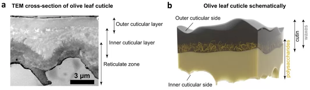
Figure 1. Olive Leaf Cuticle Structure: Cutin, Waxes, and Polysaccharides. Image reproduced from Kamtsikakis et al., 2021, Nature Communications, Figure 1a–b, licensed under CC BY 4.0.
2. Analytical Methods for Cutin and Waxes
2.1 Cuticular Wax Extraction and Profiling
Cuticular waxes are hydrophobic mixtures whose constituents differ in polarity and volatility. Their solubility in organic solvents enables extraction from intact leaves, stems, and fruits, commonly through brief contact with chloroform. This dissolves accessible wax constituents while the cutin polyester remains in the tissue. Solvent choice, exposure time, and tissue integrity influence extraction selectivity; prolonged contact or damaged surfaces can increase the recovery of internal lipids. The extract can contain both epicuticular and intracuticular waxes, depending on their accessibility under the selected conditions (Baales et al., 2021).
Gas chromatography–mass spectrometry (GC–MS) is commonly used for wax constituents that are sufficiently volatile and thermally stable, either directly or after derivatization. In conventional GC workflows, hydrocarbons are generally analyzed unchanged, while fatty acids and alcohols commonly undergo silylation to improve chromatographic behavior. Compound identification combines mass spectral matching with retention information, and gas chromatography with flame ionization detection (GC–FID) provides complementary quantitative measurements (Baales et al., 2021). Liquid chromatography–mass spectrometry (LC–MS) provides an alternative for selected high-molecular-weight or thermally labile wax esters, whose low volatility or sensitivity to heating can restrict GC analysis. Suitable ionization methods, such as atmospheric pressure chemical ionization (APCI), support their detection (Vrkoslav et al., 2010).
2.2 Cutin Depolymerization and Monomer Analysis
Cutin is a polyester that is insoluble in conventional lipid-extraction solvents. Its monomer composition is commonly characterized after depolymerization releases smaller, soluble constituents. Sample preparation generally begins with exhaustive solvent extraction to remove waxes and other extractable lipids, leaving a residue enriched in cell wall material and lipid polymers. Common depolymerization approaches include acid- and base-catalyzed methanolysis, which release fatty acid constituents as methyl esters. Catalyst concentration, temperature, and reaction duration influence cleavage efficiency and monomer recovery (Baales et al., 2021; Simões et al., 2022).
GC–MS is widely used for cutin monomers because methylation and silylation produce derivatives suitable for gas chromatographic separation. In conventional GC workflows, released monomers are recovered into an organic solvent, and their remaining hydroxyl groups are silylated. A common strategy combines GC–MS identification in representative samples with GC–FID quantification across biological replicates; calibrated GC–MS can also provide quantitative measurements (Baales et al., 2021; Bourgault & Molina, 2025). LC–MS complements this approach when the target is soluble cutin oligomers. Bhunia et al. (2018) combined partial cutinase digestion with high-resolution LC–MS to characterize dimers, trimers, and tetramers that retained information about monomer connections.
Table 2. Analytical differences between cuticular waxes and cutin
| Comparison | Cuticular wax analysis | Cutin monomer analysis |
|---|---|---|
| Analytical target | Solvent-extractable wax constituents | Products released from the insoluble polyester |
| Main preparation | Extraction; concentration; derivatization as needed | Delipidation; depolymerization; monomer recovery; derivatization |
| Typical measurements | GC–MS identification and calibrated GC-FID or GC–MS quantification | GC–MS monomer identification and calibrated GC-FID or GC–MS quantification |
| Principal outputs | Constituent amounts, class composition, and chain-length patterns | Recovered monomer amounts and composition |
| Key boundary | A solvent extract does not automatically distinguish wax layers | Monomer profiles do not reconstruct intact polymer architecture |
Method overview based on Baales et al. (2021), Jenkin and Molina (2015), and Bourgault and Molina (2025).
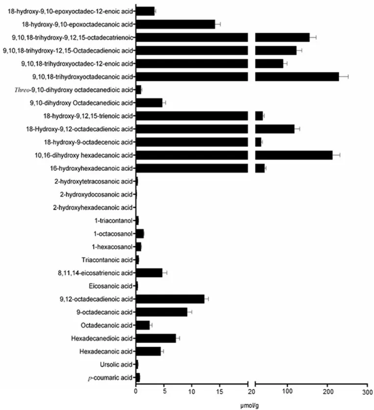
Figure 2. Apple Cutin Monomer Composition After Depolymerization. Image reproduced from Bhunia et al., 2018, Plant Methods, Figure 2, licensed under CC BY 4.0.
2.3 Cuticle Imaging and Functional Measurements
The chromatographic and mass spectrometric approaches described above characterize cutin monomers and wax constituents, including their identities and abundance. Complementary imaging methods show how the cuticle is physically organized and how its mechanical properties vary across different regions:
- Surface imaging: Scanning electron microscopy reveals wax crystals, surface texture, and cracks, providing a structural description of the surface from which waxes are extracted.
- Cross-sectional imaging: Microscopy of cuticle sections shows thickness and internal organization, adding spatial information to the chemical composition measured in the extracts.
- Mechanical mapping: Atomic force microscopy measures local properties such as stiffness. Combining these measurements with chemical imaging connects regional differences in composition with the mechanical behavior of the cuticle (Reynoud et al., 2023).
Functional measurements complement these structural observations by showing how the cuticle regulates water movement. Water-loss and permeance assays assess water transport across the cuticular barrier, whereas contact-angle measurements characterize surface wetting. Studies use isolated cuticles or controlled leaf assays that account for stomatal contributions (Shellakkutti et al., 2022; Grünhofer et al., 2024). Combining these measurements with chemical profiles connects changes in lipid composition with barrier performance and surface behavior, supporting studies of plant dehydration, fruit surface wetness, and postharvest water loss.
3. Study Design for Reliable Cuticle Lipid Analysis
3.1 Sampling and Surface-Area Normalization
Reliable cutin and wax comparisons begin with samples matched by organ, position, and developmental stage. This matters because surface chemistry changes during growth: black cottonwood leaves shift from phenolic- and terpene-rich surface profiles toward aliphatic waxes as they mature (Chen et al., 2023). Matching leaf age or fruit maturity across groups therefore helps distinguish treatment-associated changes from normal development.
Cutin and wax amounts can be expressed per unit surface area, fresh or dry tissue mass, or organ. Surface-area normalization expresses the recovered amount relative to the area covered by the cuticle. In developing strawberries, cutin and wax amounts increased per fruit but decreased per unit surface area because deposition lagged behind fruit expansion (Straube et al., 2024). Reporting amounts per square centimeter alongside organ-level totals distinguishes lipid loading per unit area from whole-organ accumulation. The area definition also needs to match the extracted surface, particularly when both leaf sides contribute to the sample.
3.2 Sample Preparation and Quality Control
Sample preparation determines how efficiently cutin monomers and wax constituents are recovered, so consistent extraction and depolymerization conditions improve comparability across samples. In cork oak leaves, increasing sodium methoxide concentration changed the proportions of released ω-hydroxyacids and dicarboxylic acids (Simões et al., 2022). Applying the same validated conditions therefore helps distinguish biological differences from variation in monomer recovery.
For quantitative comparisons, a suitable internal standard helps track processing losses and detector variation. Adding it before extraction can also help assess extraction losses when it behaves similarly to the analytes. A freely added standard provides limited information about cleavage of the native cutin polymer, so depolymerization efficiency needs separate validation. Authentic standards support compound-specific calibration, while representative standards support estimates with stated response assumptions. Procedural blanks identify background contributions, and reporting the standards and calibration basis makes quantitative results easier to compare.
4. Cutin and Wax Profile Interpretation
Cutin and wax profiles describe several different aspects of a sample: total recovered material, the contribution of chemical classes, individual compounds, and distributions such as carbon chain length. Each can change independently. Relative percentages also depend on the denominator: a compound can occupy a larger fraction of the profile because other components decreased, even if its own amount remained unchanged. Examining area-normalized amounts alongside proportions helps distinguish these possibilities and prevents a compositional shift from being mistaken for increased deposition.
The biological significance of a compositional shift becomes clearer when chemical profiles are considered alongside measured cuticle properties. In grey poplar, a cer6 mutant showed altered wax composition and approximately doubled cuticular transpiration, while total wax amount remained similar to the control (Grünhofer et al., 2024). This finding highlights the functional importance of wax composition alongside total loading. In tomato fruit, correlated chemical and mechanical imaging revealed gradients through the cuticle, with local mechanical differences associated with chemical and structural variation (Reynoud et al., 2023). Together, these studies show how combining composition, spatial organization, and physiological measurements supports a more complete interpretation of cuticle function.
5. Applications of Cuticle Lipid Analysis in Plant Research
5.1 Water-Barrier Properties and Stress Responses
Cutin and wax analysis can reveal how plant surfaces respond chemically to water limitation and whether these changes accompany altered barrier performance. In drought-adaptation research, chemical profiles help identify stress-responsive lipid classes and connect their accumulation with changes in biosynthesis and transport. In the desert shrub Ammopiptanthus mongolicus, osmotic stress increased total wax and several alkane components, accompanied by reduced epidermal permeability. Increased expression of genes involved in very-long-chain fatty acid synthesis, alkane biosynthesis, and wax transport linked these chemical changes with candidate processes contributing to surface-barrier adjustment (Sumbur et al., 2024).
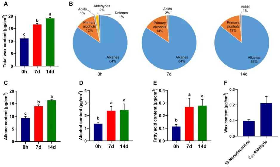
Figure 3. The effect of osmotic stress on the cuticular wax of A. mongolicus leaves. Image reproduced from Sumbur et al., 2024, Biomolecules, Figure 2A–F, licensed under CC BY 4.0.
Studies of cultivated and wild barley extended this application to both waxes and cutin. Osmotic stress increased wax amounts per unit area in most sampled leaves and cutin amounts in younger leaves, alongside denser wax crystals and increased expression of cuticle-related biosynthetic genes (Shellakkutti et al., 2022). These findings show how combined chemical and molecular profiling can characterize coordinated surface responses to water limitation. Cuticular conductance remained unchanged in barley, illustrating that the physiological effects of increased deposition depend on the plant system.
5.2 Fruit Development, Cracking, and Postharvest Quality
Cutin and wax analysis helps explain how fruit surfaces accommodate expansion and maintain integrity during development. Straube et al. (2024) found that strawberry cuticle deposition ceased around the onset of color change, while fruit expansion continued. The existing cutin and wax were consequently distributed over an increasing surface area, accompanied by greater cuticular strain and susceptibility to microcracking. These findings connected the timing of cuticle deposition with surface deterioration, helping explain why rapidly expanding strawberries become vulnerable to water soaking and cracking.
In postharvest research, cutin and wax profiles help characterize surface responses associated with quality retention. Wu et al. (2023) found that hot-water-treated Satsuma mandarins retained higher wax and cutin contents than controls during storage, while their main constituent classes remained similar. Surface wax crystals melted and redistributed, and treated fruit showed improved quality retention. These findings connect postharvest treatment with changes in cuticle abundance and surface organization, placing cuticle remodeling within the broader physiological response to heat treatment.
Plant Wax and Cutin Analysis at MetwareBio
Plant cuticular waxes and cutin contribute to water retention and the structural integrity of aerial surfaces. Understanding their composition supports research into plant stress responses, development, and fruit quality. MetwareBio offers GC–MS-based wax profiling and metabolomics methods tailored to the chemical characteristics of different cutin monomers. Our complementary plant lipidomics service profiles membrane and storage lipids, providing broader context for understanding lipid remodeling during plant growth and stress responses. Please feel free to contact us to discuss your research objectives, sample types, and suitable analytical approaches.
Contact UsRead More: Plant Lipid and Metabolomics Analysis
Cutin and cuticular waxes are part of a wider plant lipid and metabolite landscape. These related articles cover plant lipidomics workflows, metabolomics platforms, extraction methods, and fatty acid profiling that complement cuticle lipid analysis.
Profiles membrane, storage, and signaling lipids at the molecular-species level, complementing the surface lipid (cutin and wax) analysis covered in this article.
A broad entry point into plant metabolomics: metabolite classes, LC-MS/GC-MS platform selection, and study design for growth, stress, and quality questions.
Extraction solvent choice matters for internal lipids too. Compare the two dominant methods, including their behavior with plant tissues such as seeds and leaves.
Cutin monomers and wax constituents are built from long-chain fatty acid chemistry. Review fatty acid profiling platforms and chain-length analysis here.
References
- Baales, J., Zeisler-Diehl, V. V., & Schreiber, L. (2021). Analysis of extracellular cell wall lipids: Wax, cutin, and suberin in leaves, roots, fruits, and seeds. Methods in Molecular Biology, 2295, 275–293. https://doi.org/10.1007/978-1-0716-1362-7_15
- Bhunia, R. K., Showman, L. J., Jose, A., & Nikolau, B. J. (2018). Combined use of cutinase and high-resolution mass-spectrometry to query the molecular architecture of cutin. Plant Methods, 14, 117. https://doi.org/10.1186/s13007-018-0384-6
- Bonaventure, G., Beisson, F., Ohlrogge, J., & Pollard, M. (2004). Analysis of the aliphatic monomer composition of polyesters associated with Arabidopsis epidermis: Occurrence of octadeca-cis-6, cis-9-diene-1,18-dioate as the major component. The Plant Journal, 40(6), 920–930. https://doi.org/10.1111/j.1365-313X.2004.02258.x
- Bourgault, R., & Molina, I. (2025). Compositional analysis of cutin in maize leaves. Cold Spring Harbor Protocols, 2025(4), pdb.prot108434. https://doi.org/10.1101/pdb.prot108434
- Chen, J. Y., Kuruparan, A., Zamani-Babgohari, M., & Gonzales-Vigil, E. (2023). Dynamic changes to the plant cuticle include the production of volatile cuticular wax-derived compounds. Proceedings of the National Academy of Sciences, 120(49), e2307012120. https://doi.org/10.1073/pnas.2307012120
- Grünhofer, P., Herzig, L., Zhang, Q., Vitt, S., Stöcker, T., Malkowsky, Y., Brügmann, T., Fladung, M., & Schreiber, L. (2024). Changes in wax composition but not amount enhance cuticular transpiration. Plant, Cell & Environment, 47(1), 91–105. https://doi.org/10.1111/pce.14719
- Jenkin, S., & Molina, I. (2015). Isolation and compositional analysis of plant cuticle lipid polyester monomers. Journal of Visualized Experiments, 105, 53386. https://doi.org/10.3791/53386
- Kamtsikakis, A., Baales, J., Zeisler-Diehl, V. V., Vanhecke, D., Zoppe, J. O., Schreiber, L., & Weder, C. (2021). Asymmetric water transport in dense leaf cuticles and cuticle-inspired compositionally graded membranes. Nature Communications, 12, 1267. https://doi.org/10.1038/s41467-021-21500-0
- Reynoud, N., Geneix, N., D’Orlando, A., Petit, J., Mathurin, J., Deniset-Besseau, A., Marion, D., Rothan, C., Lahaye, M., & Bakan, B. (2023). Cuticle architecture and mechanical properties: A functional relationship delineated through correlated multimodal imaging. New Phytologist, 238(5), 2033–2046. https://doi.org/10.1111/nph.18862
- Shellakkutti, N., Thangamani, P. D., Suresh, K., Baales, J., Zeisler-Diehl, V., Klaus, A., Hochholdinger, F., Schreiber, L., & Kreszies, T. (2022). Cuticular transpiration is not affected by enhanced wax and cutin amounts in response to osmotic stress in barley. Physiologia Plantarum, 174(4), e13735. https://doi.org/10.1111/ppl.13735
- Simões, R., Miranda, I., & Pereira, H. (2022). Cutin extraction and composition determined under differing depolymerisation conditions in cork oak leaves. Phytochemical Analysis, 33(1), 127–135. https://doi.org/10.1002/pca.3075
- Straube, J., Hurtado, G., Zeisler-Diehl, V., Schreiber, L., & Knoche, M. (2024). Cuticle deposition ceases during strawberry fruit development. BMC Plant Biology, 24, 623. https://doi.org/10.1186/s12870-024-05327-7
- Sumbur, B., Zhou, M., Dorjee, T., Bing, J., Ha, S., Xu, X., Zhou, Y., & Gao, F. (2024). Chemical and transcriptomic analyses of leaf cuticular wax metabolism in Ammopiptanthus mongolicus under osmotic stress. Biomolecules, 14(2), 227. https://doi.org/10.3390/biom14020227
- Vrkoslav, V., Urbanová, K., & Cvačka, J. (2010). Analysis of wax ester molecular species by high performance liquid chromatography/atmospheric pressure chemical ionisation mass spectrometry. Journal of Chromatography A, 1217(25), 4184–4194. https://doi.org/10.1016/j.chroma.2009.12.048
- Wu, S., Li, X., Jiang, J., Huang, H., Cheng, X., Li, G., Shan, Y., & Zhu, X. (2023). Reveal the relationship between the quality and the cuticle composition of Satsuma mandarin (Citrus unshiu) by postharvest heat treatment. Journal of Food Science, 88(12), 4879–4891. https://doi.org/10.1111/1750-3841.16803