O-GlcNAcylation is a dynamic post-translational modification that regulates intracellular proteins involved in metabolism, signaling, transcription, stress responses, and proteostasis. Its proteome-wide analysis remains technically demanding because modified peptides are often substoichiometric, O-GlcNAc can turn over rapidly, and the glycosidic bond is labile during conventional tandem mass spectrometry. Reliable O-GlcNAc proteomics therefore depends on coordinated choices in sample preservation, enrichment, fragmentation, site localization, and quantitative interpretation. This article outlines the core enrichment strategies, LC-MS/MS approaches, site-localization considerations, and quantitative interpretation principles used in O-GlcNAcylation proteomics. It provides researchers with a practical framework for selecting an appropriate analytical workflow and interpreting O-GlcNAc changes in the context of site-level evidence and total protein abundance.
1. What Is O-GlcNAcylation?
O-linked beta-N-acetylglucosamine modification, usually called O-GlcNAcylation, is the covalent attachment of a single beta-N-acetylglucosamine residue to the hydroxyl group of serine or threonine residues on intracellular proteins. Unlike many secretory glycosylation pathways, O-GlcNAc is generally not extended into a complex glycan. Its cycling is controlled primarily by two enzymes: O-GlcNAc transferase (OGT), which transfers GlcNAc from UDP-GlcNAc to protein substrates, and O-GlcNAcase (OGA), which removes the modification. Because UDP-GlcNAc production through the hexosamine biosynthetic pathway reflects inputs from glucose, amino acid, fatty acid, and nucleotide metabolism, O-GlcNAcylation connects cellular metabolic state with protein regulation. O-GlcNAc-modified proteins are widely found in the nucleus, cytoplasm, and mitochondria, where the modification can influence protein activity, localization, interactions, turnover, transcriptional regulation, and stress adaptation. (Yang & Qian, 2017; Chatham et al., 2021)
The term O-linked glycosylation covers several chemically and biologically distinct processes, so O-GlcNAcylation should not be treated as interchangeable with classical mucin-type O-GalNAc glycosylation. Mucin-type O-glycosylation begins primarily in the secretory pathway and produces structurally diverse glycans that can be extended by multiple glycosyltransferases. Intracellular O-GlcNAcylation instead behaves more like a regulatory PTM: it is rapidly added and removed, frequently responds to nutrient and stress signals, and often interacts with phosphorylation. This distinction matters experimentally because a workflow optimized for heterogeneous secretory O-glycopeptides does not automatically provide confident O-GlcNAc site mapping. O-GlcNAc proteomics therefore requires enrichment and fragmentation strategies selected specifically for the chemical behavior and intracellular biology of this modification. (Saha et al., 2021; Maynard & Chalkley, 2021)
| Feature | Intracellular O-GlcNAcylation | Mucin-Type O-GalNAc Glycosylation |
|---|---|---|
| Initial sugar | N-acetylglucosamine (GlcNAc) | N-acetylgalactosamine (GalNAc) |
| Major modified residues | Ser/Thr | Ser/Thr |
| Typical cellular location | Nucleus, cytoplasm, mitochondria | Secretory pathway, cell surface, extracellular proteins |
| Glycan structure | Usually a single, dynamically cycling monosaccharide | Frequently extended into structurally diverse O-glycans |
| Core enzymatic regulation | OGT adds and OGA removes O-GlcNAc | GALNT enzymes initiate glycosylation, followed by additional glycosyltransferases |
| Main analytical challenge | Low/substoichiometric abundance, labile modification, site localization | Glycan microheterogeneity, glycan composition and site assignment |
| Typical proteomics objective | Identify, localize, and quantify regulatory O-GlcNAc sites | Characterize glycosites and O-glycan structures |
2. Why is O-GlcNAcylation Proteomics Challenging?
O-GlcNAcylation is more difficult to characterize than conventional protein abundance because the analytical challenge exists at several levels simultaneously. The modified peptide may represent only a small fraction of the total peptide population, the modification state can change rapidly with cellular conditions, and the O-GlcNAc linkage itself creates difficulties for residue-level identification. Together, these features affect both what can be detected and how confidently the resulting signals can be interpreted.
- Low stoichiometry and limited detectability. O-GlcNAcylation is frequently substoichiometric, meaning that only a fraction of a given protein population carries the modification at a particular site. After proteolysis, O-GlcNAc peptides can therefore be substantially less abundant than their unmodified counterparts and may be suppressed by the much larger background of unmodified peptides. Failure to detect a modified peptide consequently does not necessarily indicate that the corresponding site is unmodified; it may simply fall below the effective sampling depth of the analysis (Wang et al., 2014; Maynard & Chalkley, 2021).
- Rapid turnover and strong biological context dependence. O-GlcNAc is continuously added and removed by O-GlcNAc transferase (OGT) and O-GlcNAcase (OGA), allowing individual sites to respond rapidly to nutrient status, signaling, stress, and other cellular conditions. The measured O-GlcNAc proteome therefore represents a time- and condition-dependent regulatory state rather than a fixed property of the protein population. Differences in biological state or pre-analytical handling can consequently influence the modification profile even when total protein abundance remains relatively stable (Yang & Qian, 2017; Chatham et al., 2021).
- Labile modification and uncertain site localization. The O-glycosidic bond linking GlcNAc to Ser or Thr is relatively labile during commonly used collision-based tandem mass spectrometry. Fragmentation can therefore provide evidence that a peptide carries a HexNAc modification while losing the information needed to determine which residue was modified. This becomes particularly important when a peptide contains several candidate Ser/Thr residues, because peptide identification, detection of a modified species, and confident site localization represent different levels of analytical evidence (Maynard & Chalkley, 2021).
These challenges require specialized choices at different stages of an O-GlcNAc proteomics workflow. Low-abundance modified peptides must first be selectively captured, mass spectrometry must generate informative fragment evidence without losing critical modification information, and downstream analysis must distinguish confident site assignments from more ambiguous peptide-level detections.
3. Enrichment Strategies for O-GlcNAc Proteomics
3.1 Antibody-Based O-GlcNAc Enrichment
Antibody-based enrichment uses antibodies that recognize O-GlcNAc-containing proteins or peptides to selectively capture modified species before LC-MS/MS. Peptide-level enrichment is particularly useful for site-centric proteomics because enrichment occurs after digestion, allowing modified peptides to be introduced directly into the analytical workflow. The method is operationally straightforward and does not require chemical derivatization of the sugar.
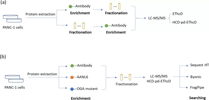
Its main limitation is selectivity bias. Antibody recognition can depend on the local peptide sequence and structural context around the modified residue, so an antibody-enriched dataset represents the subset of O-GlcNAc species efficiently recognized by the reagent. A 2024 head-to-head comparison of antibody, lectin AANL6, and an engineered OGA-based affinity reagent found substantial complementarity among the O-GlcNAc proteins and sites recovered by different enrichment methods. The result emphasizes that enrichment chemistry contributes directly to observed proteome coverage. (Hou et al., 2024)
3.2 Lectin-Based Enrichment
Lectins recognize defined carbohydrate features and provide another route to enrich O-GlcNAc species without covalently modifying the sample. Wheat germ agglutinin has historically been used in lectin weak affinity chromatography (LWAC), in which repeated weak interactions with terminal GlcNAc residues allow O-GlcNAc peptides to be separated from much of the unmodified peptide background. Newer engineered lectin reagents can provide different binding characteristics and greater selectivity.
Lectin enrichment remains dependent on carbohydrate recognition rather than direct knowledge of the modified peptide sequence, so specificity and recovery vary between reagents and sample contexts. Other GlcNAc-containing glycoconjugates can also complicate interpretation if sample preparation does not sufficiently restrict the relevant molecular population. Lectin-based enrichment is therefore best considered a distinct sampling strategy rather than a universally interchangeable substitute for antibody enrichment. (Maynard & Chalkley, 2021; Hou et al., 2024)
3.3 Chemoenzymatic Labeling and Affinity Enrichment
Chemoenzymatic methods introduce a bioorthogonal chemical handle onto O-GlcNAc residues before affinity purification. A commonly used principle employs an engineered galactosyltransferase to transfer a modified galactose derivative onto terminal O-GlcNAc. The introduced azide or related reactive handle can then be coupled through click chemistry to biotin or another affinity tag, enabling selective capture of labeled O-GlcNAc proteins or peptides.
This strategy provides a chemically defined enrichment handle and can be adapted for detection, purification, and quantitative workflows. Its increased chemical specificity comes with additional experimental steps, and overall recovery depends on enzymatic labeling efficiency, accessibility of O-GlcNAc residues, click-reaction efficiency, and subsequent affinity purification. Chemoenzymatic enrichment is therefore attractive when specificity and downstream chemical manipulation are priorities, but the additional processing must be incorporated into reproducibility and recovery assessments. (Saha et al., 2021)
3.4 How to Select an O-GlcNAc Enrichment Strategy
The appropriate enrichment strategy depends on the analytical objective rather than on a universal ranking of methods. Antibody enrichment offers a relatively direct peptide-centric workflow, lectin-based approaches provide an orthogonal carbohydrate-recognition mechanism, and chemoenzymatic labeling adds a highly manipulable affinity handle. Engineered OGA-binding reagents represent another developing strategy. Importantly, different approaches can recover partially non-overlapping O-GlcNAc populations. For discovery studies seeking broader coverage, methodological complementarity may be advantageous when sufficient material and analytical capacity are available. For comparative studies across many biological samples, a single validated enrichment workflow may provide more consistent quantitative behavior. In either case, enrichment should be assessed as part of the measurement system: reproducible capture, background contamination, site recovery, and compatibility with downstream fragmentation are more informative than enrichment yield alone. (Hou et al., 2024)
| Enrichment strategy | Capture principle | Main advantage | Main limitation | Useful context |
|---|---|---|---|---|
| Antibody-based | Recognition of O-GlcNAc-containing epitopes | Direct workflow; compatible with peptide-level enrichment | Sequence/epitope-dependent bias | Routine site-centric profiling |
| Lectin/LWAC | Carbohydrate-binding affinity | No chemical derivatization; orthogonal recognition mechanism | Weak affinity and carbohydrate cross-reactivity can influence selectivity | Broad or complementary profiling |
| Chemoenzymatic labeling | Enzymatic tagging followed by bioorthogonal affinity capture | Chemically defined enrichment handle | Additional reactions and efficiency-dependent recovery | Chemically tagged enrichment and chemical biology workflows |
| Engineered OGA affinity reagents | Binding of O-GlcNAc by modified OGA | Alternative affinity mechanism | Reagent- and workflow-dependent coverage | Complementary site discovery |
4. Mass Spectrometry Strategies for O-GlcNAc Proteomics
4.1 LC-MS/MS Detection of O-GlcNAc-Modified Peptides
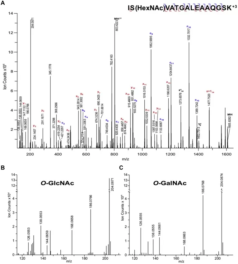
After enrichment, O-GlcNAc peptides are generally separated by liquid chromatography and analyzed by tandem mass spectrometry. High-resolution MS establishes precursor mass and isotope information, while MS/MS provides peptide-sequence and modification evidence. Collision-based spectra commonly contain the HexNAc oxonium ion near m/z 204, which is a valuable diagnostic indicator that a HexNAc-containing precursor has fragmented.
The diagnostic ion alone does not identify the modified residue, nor does it establish that the HexNAc is specifically O-GlcNAc. The analytical value of LC-MS/MS therefore depends on combining precursor evidence, peptide-backbone fragments, modification-retaining ions, enrichment context, and database-search results. For a site-specific study, an instrument can detect many glycosylated precursor signals without necessarily producing an equivalent number of confidently localized O-GlcNAc sites. (Maynard & Chalkley, 2021; Udeshi et al., 2024)
4.2 CID/HCD vs. ETD/EThcD for O-GlcNAc Site Localization
CID and HCD efficiently generate b- and y-type peptide fragments and are valuable for peptide identification and detection of HexNAc-derived diagnostic ions. Their weakness for O-GlcNAc is the preferential cleavage of the labile glycosidic bond, which can erase residue-specific modification information. Strong evidence for a HexNAc-containing peptide therefore does not guarantee strong localization evidence.
ETD fragments the peptide backbone through a different mechanism and often retains O-GlcNAc on the resulting c- and z-type ions. This preservation made ETD a major advance for site-specific O-GlcNAc proteomics. EThcD supplements ETD with collision activation, providing complementary fragment series and often richer sequence information. More recent workflows can use an HCD spectrum to recognize diagnostic HexNAc ions and trigger EThcD for the same precursor, concentrating site-informative fragmentation on likely glycopeptides. Hou et al. used such an HCD-product-dependent EThcD strategy in their systematic enrichment comparison. (Hou et al., 2024; Udeshi et al., 2024)
4.3 DDA vs. DIA Acquisition in O-GlcNAc Proteomics
Acquisition mode should be selected together with fragmentation chemistry and the required level of site resolution. DDA remains common in O-GlcNAc discovery workflows because product-dependent acquisition can identify candidate HexNAc-containing precursors and direct ETD/EThcD fragmentation toward them (Burt et al., 2022; Hou et al., 2024). DIA offers attractive quantitative completeness and consistency across sample series, but a generic DIA workflow does not automatically solve the localization problem posed by a labile modification (Lou & Shui, 2024).
For O-GlcNAc, the useful question is therefore broader than "DDA or DIA?" The workflow must provide fragment ions that distinguish candidate sites and software capable of interpreting those ions correctly. A high-throughput acquisition strategy that increases peptide detection but loses site-determining information may be suitable for some quantitative objectives yet insufficient for confident site-resolved mapping. The analytical objective should determine the balance between depth, quantitative consistency, duty cycle, and localization evidence. (Udeshi et al., 2024)
5. O-GlcNAc Site Mapping and Identification Confidence
5.1 Peptide Identification vs. Modification-Site Localization
A peptide can be confidently identified while its modification site remains ambiguous. Consider a peptide containing several Ser/Thr residues: the precursor mass establishes that a HexNAc modification is present, and backbone fragments may establish the peptide sequence, but the modification can be assigned to a specific residue only if site-determining fragment ions distinguish competing localization possibilities.
O-GlcNAc datasets should therefore report peptide identification and site localization as related but separate evidence layers. For mechanistic interpretation, the distinction is critical because mutation studies, kinase/PTM crosstalk hypotheses, structural modeling, and targeted validation all depend on knowing the correct residue. Proteome coverage expressed only as "modified peptides" or "modified proteins" cannot be interpreted as an equal number of confidently localized O-GlcNAc sites.
5.2 Distinguishing O-GlcNAc from Other HexNAc Modifications
Mass alone creates another source of ambiguity. O-GlcNAc and O-GalNAc have the same elemental composition and therefore the same nominal modification mass. The HexNAc oxonium ion is evidence for a HexNAc-containing precursor, but it cannot by itself discriminate intracellular O-GlcNAc from other HexNAc modifications.
Confidence comes from convergent evidence. Modification-specific enrichment, intracellular protein context, appropriate sample fractionation, peptide sequence and localization, characteristic fragmentation, and orthogonal biochemical validation can all narrow the interpretation. This is particularly important when extracellular or secretory glycoproteins are present in the same preparation. Database searching should not convert a generic HexNAc mass shift automatically into an O-GlcNAc biological assignment without considering this chemical ambiguity. (Wang et al., 2014; Maynard & Chalkley, 2021)
5.3 Database Searching and FDR Control
O-GlcNAc search workflows must define the relevant modification mass and candidate residues while controlling the search space sufficiently to preserve statistical discrimination. Search engines then score peptide-spectrum matches and, where supported, calculate modification-localization scores or probabilities from site-determining fragment evidence. Filtering should distinguish confidence in the peptide sequence from confidence in the residue assignment.
Software choice can influence the recovered site list. In the 2024 systematic comparison, Sequest HT, Byonic, and FragPipe produced complementary identifications from the same O-GlcNAc datasets, illustrating that computational processing is another source of methodological variation. High-value candidate sites should retain traceability to the underlying spectrum, localization evidence, search parameters, and error-control criteria. A large site table without this information is difficult to audit and can overstate biological certainty. (Hou et al., 2024)
6. Quantifying and Interpreting O-GlcNAcylation Changes
6.1 O-GlcNAc Quantification and Parent-Protein Abundance
Quantitative O-GlcNAc proteomics compares the relative abundance of modified peptides or sites across experimental conditions, but the measured signal does not directly represent modification occupancy. An increase in an O-GlcNAc peptide may reflect greater modification of a stable protein, increased abundance of the parent protein, or changes in both processes. Integrating global proteomics with O-GlcNAc proteomics provides an important reference for distinguishing PTM-level regulation from changes in overall protein abundance. Absolute site occupancy requires additional information about both modified and unmodified peptide populations and should be treated as a separate analytical objective from conventional relative PTM quantification (Olsen & Mann, 2013).
6.2 Biological Interpretation of Differential O-GlcNAc Sites
Differential O-GlcNAc sites can be mapped to protein domains, functional annotations, interaction networks, and pathway gene sets to identify biological patterns associated with the observed modification changes. Integration with phosphoproteomics can be particularly informative because O-GlcNAcylation and phosphorylation both regulate Ser/Thr residues and may interact at identical, neighboring, or functionally connected sites. These relationships are context-dependent and should not be reduced to a universal reciprocal switch. Pathway enrichment and correlated PTM changes provide evidence for candidate regulatory processes, but direct site competition, pathway activation, and functional causality require additional experimental support (Leney et al., 2017; Chatham et al., 2021).
7. How to Design an O-GlcNAc Proteomics Study
7.1 Define the Required Level of Analytical Evidence
Study design should begin with the biological question and the level of evidence needed to answer it. Broad O-GlcNAc profiling may focus on identifying and comparing modified proteins or peptides across conditions, whereas mechanistic studies often require confident residue-level localization and quantitative analysis of individual sites. This distinction influences the necessary enrichment selectivity, fragmentation strategy, localization confidence, and downstream validation. Defining the analytical endpoint before sample processing helps ensure that the workflow produces evidence at the same level as the intended biological conclusion.
7.2 Control Biological and Pre-Analytical Variation
O-GlcNAcylation responds rapidly to nutrient status, signaling, stress, and other aspects of cellular state, making experimental consistency particularly important. Treatment conditions, collection timing, sample handling, storage, extraction, biological replication, and batch organization should therefore be standardized across comparison groups. Quality control should evaluate both general LC-MS performance and the reproducibility of O-GlcNAc enrichment, because variability introduced before or during enrichment can propagate directly into quantitative results. For larger studies, pooled or reference material can help monitor analytical consistency across batches.
7.3 Match Validation to the Biological Claim
Validation requirements depend on what the discovery data are being used to claim. Confirming that a protein carries O-GlcNAc does not independently verify a specific modified residue, while confirming a site does not establish its functional consequence. Protein- or site-level findings can be followed with orthogonal biochemical assays or targeted mass spectrometry, whereas mechanistic conclusions may require site-directed mutagenesis and functional experiments examining protein activity, interaction, localization, stability, or phenotype. The validation strategy should therefore become more rigorous as the interpretation moves from association to site-specific mechanism.
8. O-GlcNAcylation Proteomics FAQ
What is the difference between O-GlcNAcylation and O-linked glycosylation?
O-linked glycosylation is a broad category of protein glycosylation in which sugars are attached to amino acid hydroxyl groups. O-GlcNAcylation is a specific intracellular PTM that adds a single GlcNAc residue to Ser/Thr sites and is dynamically regulated by OGT and OGA. In contrast, classical mucin-type O-glycosylation occurs mainly in the secretory pathway and usually produces more complex glycan structures.
Why is enrichment usually required for O-GlcNAc proteomics?
O-GlcNAc-modified peptides are often much less abundant than their unmodified counterparts in complex proteomic samples. Enrichment selectively concentrates these modified peptides before LC-MS/MS, improving their detectability and increasing the number of O-GlcNAc peptides and sites that can be characterized. Common approaches include antibody-based enrichment, lectin affinity, and chemoenzymatic labeling.
Why are ETD and EThcD useful for O-GlcNAc site mapping?
ETD and EThcD help preserve the labile O-GlcNAc modification while fragmenting the peptide backbone. This generates site-informative fragment ions that can distinguish among multiple candidate Ser/Thr residues within the same peptide. EThcD adds supplemental collisional activation to ETD, often providing richer sequence coverage and improving confidence in O-GlcNAc site localization.
Can LC-MS/MS distinguish O-GlcNAc from other HexNAc modifications?
LC-MS/MS can support confident O-GlcNAc assignment when site-localizing fragmentation is combined with appropriate O-GlcNAc enrichment and biological context. O-GlcNAc and O-GalNAc have the same elemental composition, so precursor mass alone cannot distinguish them. Reliable identification therefore relies on the combined evidence from enrichment specificity, peptide sequence, fragmentation behavior, and modification-site localization.
Planning an O-GlcNAcylation Proteomics Project with MetwareBio
O-GlcNAcylation studies require an analytical strategy that can capture modification-specific changes with appropriate site-level evidence. MetwareBio provides PTM proteomics services based on modification-specific enrichment, LC-MS/MS analysis, site-level identification and quantification, and downstream bioinformatics, with integrated proteome + PTM analysis available when total protein abundance is also relevant to interpretation. If you are planning to investigate O-GlcNAcylation in your research, please contact us to discuss your project requirements and a suitable analytical strategy.
Contact UsPTM Proteomics Service
The service hub for site-resolved modification profiling — modification-specific enrichment, high-resolution LC-MS/MS, confident site localization, and the differential and functional interpretation that follows an O-GlcNAc experiment.
Proteome + PTM Analysis
Why O-GlcNAc signals need a parent-protein reference: this combined workflow quantifies global protein abundance and PTM sites in parallel from the same extract, separating modification-level regulation from protein-level changes.
Phosphoproteomics in Action: Strategies for Investigating Protein Phosphorylation
A companion PTM workflow from sample preparation through TiO₂/IMAC enrichment, DDA vs DIA acquisition, and site identification — useful context for the O-GlcNAc/phosphorylation crosstalk discussed in section 6.2.
Phosphoproteomics in Drug Discovery: Revealing Hidden Pathways for Targeted Therapies
Shows how enriched PTM peptides are handled end to end, including enrichment strategy selection and site-localization scoring tools such as Ascore and ptmRS — directly comparable to the O-GlcNAc localization problem.
N-Glycosylation Proteomics
A glycosylation-focused counterpart: HILIC-based glycopeptide enrichment with 4D label-free LC-MS/MS for site-level glycosylation profiling, illustrating how enrichment chemistry shapes which glycosylated species are recovered.
An Overview of Mainstream Proteomics Techniques
Background on label-free DDA and DIA acquisition, TMT/iTRAQ labeling, and the trade-offs between depth and quantitative consistency — the same acquisition decisions that section 4.3 raises for O-GlcNAc proteomics.
References
- Burt, R. A., Alghusen, I. M., John Ephrame, S., Villar, M. T., Artigues, A., & Slawson, C. (2022). Mapping the O-GlcNAc modified proteome: Applications for health and disease. Frontiers in Molecular Biosciences, 9, 920727. https://doi.org/10.3389/fmolb.2022.920727
- Chatham, J. C., Zhang, J., & Wende, A. R. (2021). Role of O-linked N-acetylglucosamine protein modification in cellular (patho)physiology. Physiological Reviews, 101(2), 427–493. https://doi.org/10.1152/physrev.00043.2019
- Hou, C., Wu, C., Wu, Z., Cheng, Y., Li, W., Sun, H., & Ma, J. (2024). Systematic evaluation of affinity enrichment methods for O-GlcNAc proteomics. Journal of Proteome Research, 23(10), 4422–4432. https://doi.org/10.1021/acs.jproteome.4c00388
- Leney, A. C., El Atmioui, D., Wu, W., Ovaa, H., & Heck, A. J. R. (2017). Elucidating crosstalk mechanisms between phosphorylation and O-GlcNAcylation. Proceedings of the National Academy of Sciences of the United States of America, 114(35), E7255–E7261. https://doi.org/10.1073/pnas.1620529114
- Lou, R., & Shui, W. (2024). Acquisition and Analysis of DIA-Based Proteomic Data: A Comprehensive Survey in 2023. Molecular & Cellular Proteomics, 23(2), 100712. https://doi.org/10.1016/j.mcpro.2024.100712
- Maynard, J. C., & Chalkley, R. J. (2021). Methods for enrichment and assignment of N-acetylglucosamine modification sites. Molecular & Cellular Proteomics, 20, 100031. https://doi.org/10.1074/mcp.R120.002206
- Olsen, J. V., & Mann, M. (2013). Status of large-scale analysis of post-translational modifications by mass spectrometry. Molecular & Cellular Proteomics, 12(12), 3444–3452. https://doi.org/10.1074/mcp.O113.034181
- Saha, A., Bello, D., & Fernandez-Tejada, A. (2021). Advances in chemical probing of protein O-GlcNAc glycosylation: Structural role and molecular mechanisms. Chemical Society Reviews, 50(18), 10451–10485. https://doi.org/10.1039/D0CS01275K
- Udeshi, N. D., Hart, G. W., & Slawson, C. (2024). From fringe to the mainstream: How ETD MS brought O-GlcNAc to the masses. Molecular & Cellular Proteomics, 23(11), 100859. https://doi.org/10.1016/j.mcpro.2024.100859
- Wang, S., Yang, F., Camp, D. G. II, Rodland, K., Qian, W.-J., Liu, T., & Smith, R. D. (2014). Proteomic approaches for site-specific O-GlcNAcylation analysis. Bioanalysis, 6(19), 2571–2580. https://doi.org/10.4155/bio.14.239
- Yang, X., & Qian, K. (2017). Protein O-GlcNAcylation: Emerging mechanisms and functions. Nature Reviews Molecular Cell Biology, 18(7), 452–465. https://doi.org/10.1038/nrm.2017.22