G protein-coupled receptors (GPCRs) are seven-transmembrane receptors that convert extracellular cues into intracellular signals. A shared membrane architecture allows the family to respond to light, odors, neurotransmitters, hormones, lipids, metabolites, peptides, and proteins, yet generate responses that differ in strength, duration, location, and cellular effect. This versatility reflects dynamic receptor conformations, selective transducer coupling, and regulation by trafficking and cellular context. These same properties have made GPCRs central to modern pharmacology. This article follows the path from seven-transmembrane structure and G-protein activation to downstream signaling, druggability, and mass spectrometry/proteomics approaches for studying receptor dynamics and signaling networks.
1. GPCR Structure and Families: The Seven-Transmembrane Receptor Superfamily
GPCRs share a compact seven-transmembrane core, but they are not structurally or pharmacologically uniform. Different families adapt this common framework to distinct extracellular domains, ligands, and activation mechanisms.
1.1 Seven-Transmembrane GPCR Structure and Topology
A canonical GPCR is a single polypeptide with an extracellular N-terminus, an intracellular C-terminus, and seven hydrophobic transmembrane alpha-helices (TM1-TM7). Three extracellular and three intracellular loops connect the helices. Extracellular regions contribute to ligand access and recognition, whereas intracellular loops and the cytoplasmic ends of the helices form interfaces for G proteins and regulatory proteins. Many receptors also contain a membrane-associated helix 8.
The shorthand “7TM receptor” is useful but not perfectly synonymous with GPCR. Some proteins contain seven transmembrane segments without signaling through heterotrimeric G proteins, so topology alone does not establish functional classification (Alexander et al., 2025). Figure 1 summarizes this shared membrane topology.
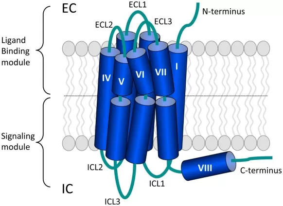
Figure 1. Seven-transmembrane topology of a representative class A GPCR. Image reproduced from de Munnik et al. (2015), Frontiers in Pharmacology, licensed under CC BY 4.0.
1.2 Major GPCR Families and Ligand Diversity
The human GPCR repertoire includes around 800 receptors, with roughly half associated with sensory functions and hundreds of non-sensory receptors regulating physiology across major organ systems (Alexander et al., 2025). Major branches include Class A/rhodopsin-like, Class B1/secretin, adhesion GPCRs, Class C/glutamate, and Class F/Frizzled receptors, which share a 7TM core but differ in extracellular architecture and ligand-recognition strategies.
GPCR diversity also connects the family directly to metabolite biology. GABA-B receptors, the bile acid receptor GPBAR1/TGR5, and free fatty acid receptors such as the short-chain fatty acid-responsive FFAR2 and FFAR3 illustrate how neurotransmitters and metabolites are sensed at the cell surface and translated into downstream responses.
2. How GPCR Structure Converts Ligand Binding into Receptor Activation
Ligand binding and G-protein engagement occur on opposite sides of the membrane. GPCR activation is therefore an allosteric process in which ligand-dependent changes propagate through the transmembrane bundle to reshape the intracellular signaling interface.
2.1 Ligand Recognition and 7TM Activation
Endogenous ligands commonly bind an orthosteric site, whereas allosteric ligands bind elsewhere and alter affinity, efficacy, or signaling behavior. Because ligand binding and G-protein engagement occur on opposite sides of the membrane, activation depends on long-range allosteric communication through residue contacts, hydration networks, ions, and helix packing. Allosteric sites can therefore provide pharmacological control without directly competing with an endogenous ligand and may offer subtype selectivity when orthosteric pockets are highly conserved. The effect of an allosteric ligand can also depend on which orthosteric ligand is present, so its pharmacology is often best interpreted as a property of a receptor-ligand system rather than of the compound alone.
Class A GPCRs provide the best-studied structural framework. Agonist binding is commonly associated with outward movement of the cytoplasmic end of TM6, rearrangements in TM5 and TM7, and rewiring of conserved networks involving motifs such as DRY, PIF, CWxP, NPxxY, and the sodium-binding pocket. Together, these changes create an intracellular cavity that accommodates the C-terminal α5 helix of Gα (Zhou et al., 2019).
These microswitches summarize a shared Class A activation logic rather than a universal checklist for every GPCR. The broader principle is coordinated communication from the ligand-binding region through the 7TM bundle to the cytoplasmic transducer interface; other GPCR classes use related allosteric principles but different structural routes. Figure 2 illustrates this representative Class A model.
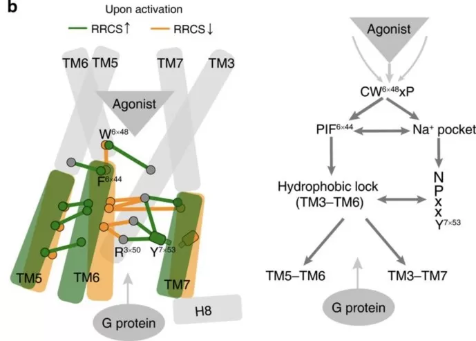
Figure 2. Representative class A GPCR activation model linking agonist binding, conserved microswitches, and G-protein coupling. Image cropped from Figure 5b in Zhou et al. (2019), eLife, licensed under CC BY 4.0; panel b only, with no scientific alteration.
2.2 GPCR Conformational Ensembles and Dynamic Signaling States
Inactive and active structures are useful reference states, but GPCRs populate broader conformational ensembles whose distributions shift with ligand chemistry, membrane environment, receptor sequence, ions, and interacting proteins. A ligand therefore changes the probability of occupying particular states rather than forcing a receptor into one universal active conformation. Static X-ray and cryo-EM structures remain essential, but each captures only part of this dynamic landscape.
This ensemble view helps explain differences in efficacy and signaling preference. A partial agonist may enrich some active-like states without stabilizing the full set associated with efficient transducer activation, while another ligand may favor states with different G-protein or arrestin compatibility. For drug discovery, the key question is not only whether a ligand binds, but which receptor states it favors and how those states are interpreted in a particular cellular context.
3. How GPCRs Activate Heterotrimeric G Proteins
Once an activated GPCR engages a heterotrimeric G protein, it promotes GDP release from Gα and initiates the nucleotide-exchange cycle. Which G protein is recruited, and how efficiently it is activated, depends on the receptor, ligand, G-protein subtype, and cellular context.
3.1 The Heterotrimeric G-Protein Activation Cycle
Heterotrimeric G proteins contain Gα, Gβ, and Gγ subunits. In the resting state, Gα is generally GDP-bound and associated with Gβγ at the cytoplasmic face of the membrane. Gα proteins are grouped into Gs, Gi/o, Gq/11, and G12/13 families, while Gβγ can also regulate ion channels, enzymes, and kinase pathways after receptor activation. Membrane anchoring of the subunits keeps the heterotrimer close to receptors and effectors, helping organize rapid signal transfer at the cytoplasmic membrane surface.
An agonist-stabilized GPCR engages the C-terminal α5 helix of Gα and promotes rearrangements that destabilize GDP. GTP binding then produces an active Gα-GTP state and a functional rearrangement or separation of Gα and Gβγ, allowing both components to regulate effectors. Gα GTPase activity, often accelerated by RGS proteins, hydrolyzes GTP and favors system reset. Time-resolved cryo-EM of the β2-adrenergic receptor-Gs complex captured sequential intermediates after GTP addition, showing coordinated changes in the nucleotide pocket, switch regions, α5 helix, and receptor interface rather than a single instantaneous transition (Papasergi-Scott et al., 2024).
3.2 G-Protein Coupling Selectivity and Assay Context
A GPCR should not automatically be assigned to only one G-protein family. Large-scale coupling maps show a spectrum from strong selectivity to broad promiscuity, and apparent profiles can differ across assay platforms (Hauser et al., 2022). Preferred coupling therefore describes a relative tendency under defined conditions, not an exclusive rule.
The readout also matters. Recruitment biosensors, nucleotide-exchange assays, and cAMP or calcium measurements interrogate different steps of the signaling sequence and are influenced differently by receptor expression and signal amplification. A ligand can therefore alter transducer coupling without producing an equivalent change in a distal cellular phenotype.
4. GPCR Downstream Signaling: Second Messengers, β-Arrestins, and Compartmentalization
After G-protein activation, signaling quickly branches through multiple effectors, second messengers, and regulatory proteins. Signal amplitude, duration, and location are further shaped by feedback, degradation, scaffolding, and receptor trafficking.
4.1 Canonical G-Protein Signaling Pathways
The four major Gα families establish different routes into the signaling network, while Gβγ adds parallel regulation of channels and kinases. Early outputs can converge on ERK/MAPK, PI3K-AKT, transcriptional programs, cytoskeletal pathways, and metabolic enzymes. The major Gα families and their canonical outputs are summarized in Table 1.
| Gα family | Representative effector | Typical output | Interpretation note |
|---|---|---|---|
| Gs | Adenylyl cyclase | Increased cAMP; PKA | Depends on receptor reserve and phosphodiesterase activity |
| Gi/o | Adenylyl cyclase; ion-channel regulators | Reduced cAMP; Gβγ signaling | Not simply the “opposite” of Gs |
| Gq/11 | PLCβ | IP₃, DAG, Ca²⁺, PKC | Strongly time- and compartment-dependent |
| G12/13 | RhoGEFs | RhoA; cytoskeletal/transcriptional responses | Less uniform across receptor systems |
4.2 Signal Amplification, Crosstalk, and Cellular Context
Second messengers such as cAMP and Ca²⁺ can be amplified, degraded, sequestered, or generated in restricted cellular regions. Phosphodiesterases, kinases, phosphatases, scaffolds, and ion channels create feedback and crosstalk, so pathway output depends on more than the receptor and G-protein family. For example, cAMP can be restricted to local microdomains, while Ca²⁺ responses are shaped by release, buffering, sequestration, and extrusion.
These regulatory layers help explain why the same receptor-ligand pair can produce different amplitudes or durations in different cells. Receptor reserve, effector abundance, organelle organization, and scaffold proteins all influence the response; receptor mRNA or total protein abundance is therefore context, not a direct proxy for signaling strength.
4.3 β-Arrestins, Trafficking, and Compartmentalized Signaling
Activated receptors are often phosphorylated by GPCR kinases (GRKs), which can favor β-arrestin recruitment, reduce further G-protein coupling, and promote internalization. Internalized receptors may recycle to the membrane, be directed toward degradation, or in some cases remain signaling competent from endosomes. For the β2-adrenergic receptor, endosome-localized Gαs can support a local RAF-MEK-ERK axis that propagates to nuclear responses (Kwon et al., 2022). The phosphorylation pattern on the receptor can influence arrestin engagement and trafficking outcome, adding another layer of receptor-specific regulation.
β-arrestin functions and signaling bias are receptor- and context-dependent. Arrestins can organize regulatory or signaling complexes, while ligands may differ in preference for G-protein families, β-arrestin recruitment, signaling compartments, or kinetics. Such bias must be defined relative to a reference ligand, pathway, and experimental system. Figure 3 places these processes in their spatial and trafficking context.
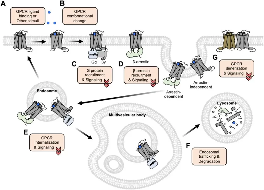
Figure 3. GPCR activation, transducer recruitment, internalization, and compartmentalized signaling; dimerization is shown as an additional regulatory mode. Image reproduced from Kim et al. (2022), Frontiers in Cell and Developmental Biology, licensed under CC BY 4.0.
5. Why GPCRs Are Major Drug Targets
GPCRs combine biological importance with several features that favor drug development, including accessible binding sites, multiple modes of pharmacological modulation, signal amplification, mature functional assays, and extensive clinical precedent.
5.1 Why GPCRs Are Highly Druggable and Pharmacologically Versatile
Many GPCR binding regions are accessible from the extracellular space or membrane-facing cavities, and endogenous ligands provide pharmacological starting points. Orthosteric and allosteric sites, receptor subtypes, tissue distribution, signaling selectivity, and compartment-specific responses offer multiple ways to tune activity and improve therapeutic selectivity. The family also supports mature functional assays for cAMP, calcium, β-arrestin recruitment, trafficking, and G-protein activation, although a strong in vitro response or receptor occupancy does not by itself establish the desired effect in tissue or patients.
GPCR pharmacology also extends beyond simple agonism and antagonism. Ligands can vary independently in efficacy, binding site, signaling preference, and molecular modality: full or partial agonists, inverse agonists, allosteric modulators, biased or bitopic ligands, and small molecules, peptides, or antibodies describe different dimensions of a pharmacological profile. Keeping these dimensions separate makes compounds easier to compare and avoids treating one assay readout as a complete description of mechanism. This flexibility is one reason the same receptor can support very different discovery strategies, from classical orthosteric screening to allosteric or signaling-selective programs.
5.2 A Mature but Underexploited GPCR Target Landscape
A 2025 GPCR drug-discovery resource catalogued 516 approved drugs acting through GPCRs, corresponding to 36% of approved drugs in that analysis, but covering only 121 GPCR targets (Caroli et al., 2025). The family therefore has extensive clinical validation while many non-sensory and orphan receptors remain underexplored.
Important challenges remain: conserved orthosteric pockets can limit subtype selectivity, receptor reserve and tissue expression can alter apparent pharmacology, responses may differ across species, and pathway preference can shift with cellular context. Productive drug discovery increasingly depends on linking a defined receptor state and signaling mechanism to the intended phenotype, not simply finding a molecule that binds.
6. Mass Spectrometry and Proteomics for GPCR Dynamics, Interactions, and Signaling
X-ray crystallography and cryo-EM define GPCR architecture in remarkable detail, but each structure captures only part of a dynamic system. Conformational exchange, transient receptor-transducer complexes, and rapid remodeling of cellular signaling networks require complementary methods such as mass spectrometry and proteomics.
6.1 HDX-MS and Covalent Footprinting for GPCR Conformational Dynamics
Hydrogen-deuterium exchange mass spectrometry (HDX-MS) reports local solvent accessibility and structural dynamics. Comparing ligand conditions can identify regions whose protection or flexibility changes with agonists, partial agonists, antagonists, or allosteric modulators. A 2024 β1-adrenergic receptor study analyzed nine ligands and identified ligand-dependent dynamic fingerprints across the receptor (Toporowska et al., 2024).
HDX-MS usually provides peptide-level rather than atomic resolution, and membrane-protein coverage and lipid/detergent conditions can affect results. Covalent footprinting can add complementary accessibility information, but dynamic differences still require functional validation. Figure 4 shows representative HDX-MS readouts for conformational changes across both tβ1AR and miniGs within an activated receptor-transducer complex.
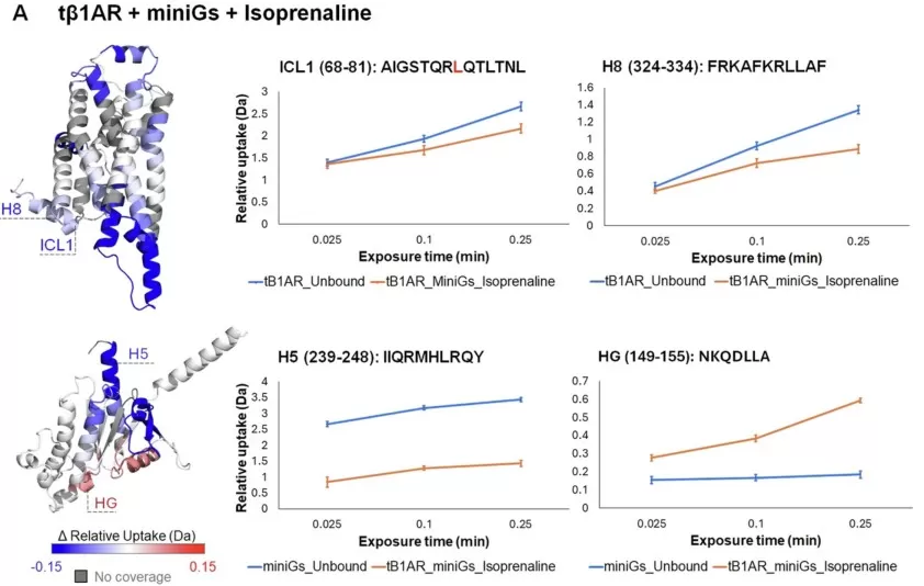
Figure 4. HDX-MS mapping of conformational changes in the isoprenaline-bound tβ1AR-miniGs complex, highlighting receptor ICL1/H8 and miniGs H5/HG. Image reproduced from Figure 6A in Toporowska et al. (2024), Nature Communications, licensed under CC BY 4.0.
6.2 Receptor-Transducer Complexes and GPCR Interactomes
Native MS can preserve noncovalent receptor-ligand-transducer assemblies under controlled conditions and has been used with β1-adrenergic receptor complexes to connect ligand binding with distinct mini-G-protein complexes and biased signaling (Yen et al., 2022). Cross-linking MS (XL-MS) adds distance restraints for integrative modeling; studies of the activated GLP-1 receptor-Gs complex revealed conformational ensembles beyond a single cryo-EM state (Yuan et al., 2023). These approaches provide structural constraints rather than complete cellular context.
For cellular interaction networks, affinity purification-MS favors relatively stable receptor-associated complexes, whereas proximity labeling with BioID, TurboID, or APEX2 can capture weak, transient, or compartment-specific proximal proteins. A 2025 APEX2 study of the luteinizing hormone receptor mapped rapid agonist-dependent changes in its local protein environment (Shchepinova et al., 2025). Proximity does not prove direct binding, so candidate proteins still require orthogonal validation. Together, these methods extend structural analysis from purified receptor complexes toward the transient and spatially organized protein environments that shape signaling in cells.
6.3 Quantitative Proteomics and Phosphoproteomics for GPCR Signaling
Quantitative proteomics can profile signaling proteins, regulators, and effectors, whereas phosphoproteomics provides a more activity-sensitive view of kinase and phosphatase networks. Time-resolved designs can reveal ligand-specific phosphosignatures involving GRKs, arrestins, MAPK, AKT, PKC, and other signaling regulators.
A differential phosphosite is not automatically a direct consequence of the target GPCR. Antagonists, receptor perturbation, rescue experiments, matched time courses, and orthogonal second-messenger assays help establish receptor dependence. Proteomics combined with metabolomics can further connect signaling-network changes with metabolic phenotypes, while Table 2 summarizes the main MS and proteomics approaches and their interpretive limits.
| Research question | Recommended approach | Primary readout | Main limitation |
|---|---|---|---|
| Which receptor regions change dynamics after ligand binding? | HDX-MS; covalent footprinting | Protection/exposure changes | Peptide-level resolution; variable membrane coverage |
| Which ligands favor particular receptor-G-protein complexes? | Native MS | Complex composition; relative coupling | Purified context |
| What conformations occur in transient GPCR complexes? | XL-MS + integrative modeling | Distance restraints; ensembles | Requires modeling and orthogonal validation |
| Which proteins appear near an activated/internalized receptor? | APEX2, TurboID, BioID | Proximal proteins | Proximity does not prove direct binding |
| Which stable proteins form receptor-associated complexes? | AP-MS | Enriched partners | Transient interactions may be lost |
| Which pathways change after activation? | Quantitative phosphoproteomics | Differential phosphosites; pathway-level signaling signatures | Direct and indirect effects must be separated |
| Does signaling reshape metabolism? | Proteomics + metabolomics | Protein and metabolite pathway changes | Correlation does not establish receptor control |
6.4 Integrating Structural, Proteomic, and Functional Evidence
A convincing GPCR mechanism usually emerges from complementary evidence. Receptor abundance and surface localization establish presence and accessibility; binding or target-engagement assays test ligand engagement; structural or biophysical measurements examine conformation; transducer assays measure G-protein or arrestin engagement; and second-messenger or phosphoproteomic readouts characterize pathway propagation. Genetic perturbation, rescue, and well-controlled pharmacology are then used to strengthen causal links to the phenotype. The strongest interpretation comes from linking these layers in the same biological model and at time points appropriate to the signaling event being tested.
These measurements are complementary rather than interchangeable. No single readout, from receptor abundance to a phosphoproteomic change, is sufficient to establish the complete mechanism or its clinical relevance.
7. How MetwareBio Supports GPCR Signaling and Drug-Response Research
GPCR studies often need molecular context beyond a receptor-specific assay. MetwareBio supports this layer with DIA quantitative proteomics, phosphoproteomics, Proteome + PTM Analysis, and integrated proteomics-metabolomics workflows for measuring signaling proteins, phosphorylation-network responses, and downstream metabolic phenotypes. These omics data are most informative when interpreted alongside receptor-specific binding, coupling, trafficking, and functional experiments matched to the biological question.
Studying GPCR-regulated signaling or drug response? MetwareBio can help design a proteomics, phosphoproteomics, or integrated multi-omics workflow that links receptor-associated molecular changes to downstream pathways and phenotypes.
Contact UsRead More: GPCR Signaling and Proteomics
These articles cover the proteomics and multi-omics methods that complement GPCR structural biology, from quantitative proteomics and phosphoproteomics to multi-omics integration for signaling network analysis.
Overview of DIA quantitative proteomics for measuring signaling proteins and effectors in GPCR studies, providing reproducible deep proteome coverage for drug-response research.
Phosphoproteomics platform for profiling kinase and phosphatase network responses after GPCR activation, capturing ligand-specific phosphosignatures at pathway level.
How integrated proteomics and metabolomics workflows connect signaling-network changes with downstream metabolic phenotypes in receptor and drug-response studies.
Combined proteomics and post-translational modification analysis services, supporting phosphorylation, ubiquitination, acetylation, and other PTM profiling for signaling research.
Comparison of DDA and DIA acquisition modes for quantitative proteomics, helping select the appropriate platform for signaling protein detection and drug-response profiling.
Methods for integrating multi-omics data layers, connecting proteomic, metabolomic, and phosphoproteomic changes to build mechanistic models of receptor signaling.
References
- Alexander, S. P. H., Davenport, A. P., Kelly, E., et al. (2025). The Concise Guide to PHARMACOLOGY 2025/26: G protein-coupled receptors. British Journal of Pharmacology, 182(S1), S24-S151. https://doi.org/10.1111/bph.70230
- Caroli, J., Andreassen, S. N., Sánchez Lorente, J., et al. (2025). An online GPCR drug discovery resource. npj Drug Discovery, 2, 17. https://doi.org/10.1038/s44386-025-00010-9
- de Munnik, S. M., Smit, M. J., Leurs, R., & Vischer, H. F. (2015). Modulation of cellular signaling by herpesvirus-encoded G protein-coupled receptors. Frontiers in Pharmacology, 6, 40. https://doi.org/10.3389/fphar.2015.00040
- Hauser, A. S., Avet, C., Normand, C., et al. (2022). Common coupling map advances GPCR-G protein selectivity. eLife, 11, e74107. https://doi.org/10.7554/eLife.74107
- Kim, H., Baek, I.-Y., & Seong, J. (2022). Genetically encoded fluorescent biosensors for GPCR research. Frontiers in Cell and Developmental Biology, 10, 1007893. https://doi.org/10.3389/fcell.2022.1007893
- Kwon, Y., Mehta, S., Clark, M., et al. (2022). Non-canonical beta-adrenergic activation of ERK at endosomes. Nature, 611, 173-179. https://doi.org/10.1038/s41586-022-05343-3
- Papasergi-Scott, M. M., Pérez-Hernández, G., Batebi, H., et al. (2024). Time-resolved cryo-EM of G-protein activation by a GPCR. Nature, 629, 1182-1191. https://doi.org/10.1038/s41586-024-07153-1
- Shchepinova, M. M., Richardson, R., Houghton, J. W., et al. (2025). Spatiotemporally resolved GPCR interactome uncovers unique mediators of receptor agonism. Cell Chemical Biology, 32(5), 722-735.e7. https://doi.org/10.1016/j.chembiol.2025.04.006
- Toporowska, J., Kapoor, P., Musgaard, M., et al. (2024). Ligand-induced conformational changes in the β1-adrenergic receptor revealed by hydrogen-deuterium exchange mass spectrometry. Nature Communications, 15, 8993. https://doi.org/10.1038/s41467-024-53161-0
- Yen, H.-Y., Liko, I., Song, W., et al. (2022). Mass spectrometry captures biased signalling and allosteric modulation of a G protein-coupled receptor. Nature Chemistry, 14, 1375-1382. https://doi.org/10.1038/s41557-022-01041-9
- Yuan, S., Xia, L., Wang, C., et al. (2023). Conformational dynamics of the activated GLP-1 receptor-Gs complex revealed by cross-linking mass spectrometry and integrative structure modeling. ACS Central Science, 9(5), 992-1007. https://doi.org/10.1021/acscentsci.3c00063
- Zhou, Q., Yang, D., Wu, M., et al. (2019). Common activation mechanism of class A GPCRs. eLife, 8, e50279. https://doi.org/10.7554/eLife.50279