Membrane proteins and other poorly soluble proteins are often under-represented in bottom-up proteomics because hydrophobicity, lipid association, aggregation, and limited protease accessibility reduce both protein recovery and peptide detectability. Reliable analysis therefore depends on the full sample-preparation workflow—from enrichment and solubilization to detergent removal, digestion, and LC-MS/MS—rather than on extraction yield alone. This article examines how these steps can be combined to improve recovery, reduce losses during digestion and cleanup, and determine whether higher total protein counts translate into better membrane-specific coverage.
1. Why Membrane and Insoluble Proteins Are Underrepresented in Proteomics
The term “membrane protein” describes a biological localization or mode of membrane association, whereas “insoluble protein” describes behavior under a particular extraction condition. Integral membrane proteins are embedded in the lipid bilayer. Many span the membrane through one or more alpha-helices, whereas beta-barrel transmembrane proteins form a distinct structural class. Multi-pass transmembrane proteins can be especially hydrophobic and analytically challenging. Peripheral membrane proteins and lipid-anchored proteins can associate strongly with membranes without sharing the same transmembrane architecture. Poorly soluble material can also include cytoskeletal and extracellular-matrix proteins, organelle-associated components, or aggregates from large protein complexes. An insoluble fraction is therefore not equivalent to a membrane fraction, and membrane-associated proteins do not all behave the same way during extraction.
Loss can occur at several stages. Incomplete membrane disruption leaves material unextracted, while centrifugation can place target proteins in a pellet that is later discarded. Detergent removal or precipitation can drive re-aggregation, and digestion of hydrophobic regions may yield peptides that remain difficult to solubilize, chromatograph, or ionize. Residual lipids, salts, and surfactants can then suppress downstream LC-MS/MS performance. These are distinct sources of bias, which is why successful protein extraction does not guarantee successful peptide identification (Masuda et al., 2021; Jiang et al., 2024).
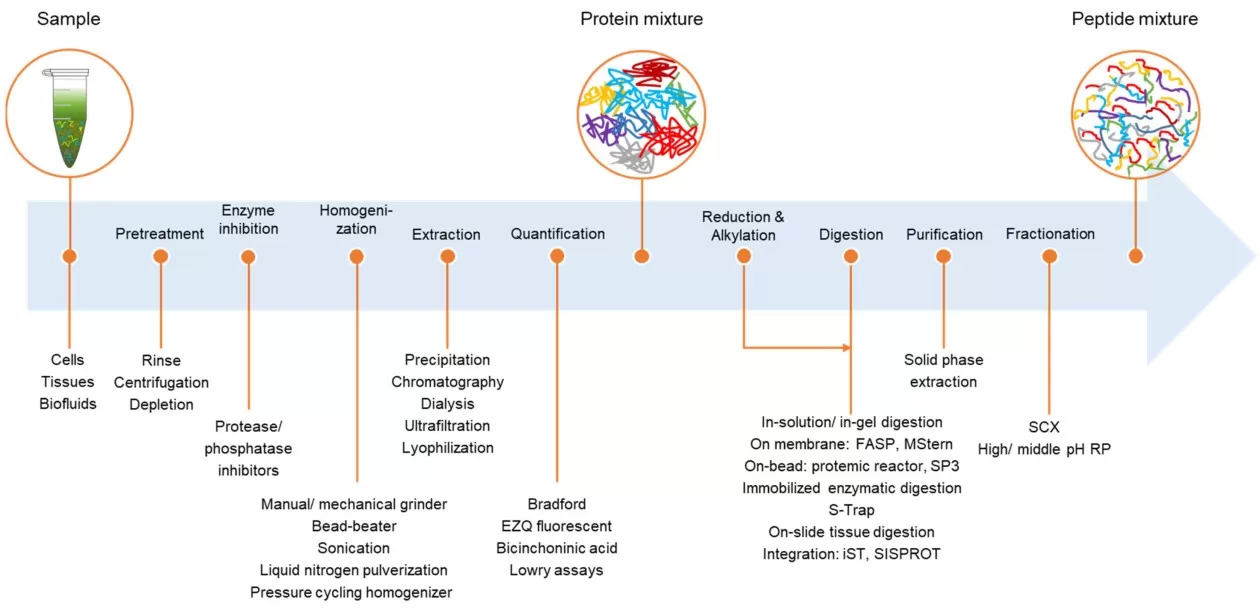
Figure 1. General sample-preparation workflow for bottom-up proteomics, from sample pretreatment and protein extraction to reduction/alkylation, digestion, purification, and peptide fractionation. Reproduced from Figure 1 in Duong and Lee (2023), International Journal of Molecular Sciences, under CC BY 4.0.
2. Membrane Protein Sample Preparation: Analytical Goal and Sample Matrix
The extraction strategy depends first on what must be preserved in the sample. Projects that require native activity, ligand binding, intact complexes, or affinity capture generally favor milder nonionic or zwitterionic detergents because extensive denaturation would compromise those readouts. In contrast, bottom-up identification and relative quantification can tolerate stronger denaturing conditions, provided that detergent removal, buffer exchange, and protease compatibility are planned from the start (Behnke & Urner, 2023).
Sample matrix determines which preparation problem dominates. Lipid-rich tissues carry more lipids into extraction and cleanup, increasing the risk of phase-separation problems and ion suppression; fibrous tissues and plant material often require more effective mechanical disruption; and organelle-enriched preparations reduce complexity but make fraction purity and cross-contamination more important. It is therefore useful to decide early whether the goal is broad membrane-proteome coverage or a defined compartment. For compartment-specific studies, subcellular fractionation usually provides more selectivity than simply increasing detergent strength (Masuda et al., 2021).
3. Membrane Protein Enrichment, Fractionation, and Sequential Extraction
Fractionation is particularly useful when membrane proteins are diluted by a much larger soluble proteome. Differential centrifugation can separate nuclei and large debris from lighter membrane-containing fractions, while ultracentrifugation or organelle-oriented protocols can generate crude membrane, microsomal, mitochondrial, or other subcellular preparations. Fractionation reduces soluble-protein dominance and increases the relative representation of the target membrane fraction before digestion. Subcellular enrichment is a common membrane-proteomics strategy, and experimental studies show that membrane washing and fractionation can increase detection of integral and multi-pass transmembrane proteins (Masuda et al., 2021; Kongpracha et al., 2022).
3.1 Membrane Washing for Membrane Protein Enrichment: Benefits and Trade-Offs
High-salt, urea, or alkaline/carbonate washes can reduce soluble or loosely associated contaminants before membrane solubilization. This may improve the relative abundance of integral membrane proteins, but stronger washing can also remove peripheral proteins or disturb weak protein-protein interactions. Kongpracha et al. (2022), for example, showed that urea- and alkaline-washed membrane fractions changed the representation of transmembrane proteins and that the choice of wash affected the retained interaction landscape. In practice, wash conditions should remove enough soluble contamination to improve membrane-protein representation without unnecessarily stripping peripheral proteins or weak complexes.
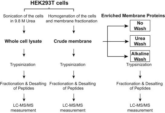
Figure 2. Membrane fractionation and washing for membrane proteomic analysis. Whole-cell lysate and crude membrane preparations were compared with no-wash, urea-wash, and alkaline-wash conditions before tryptic digestion, peptide cleanup, and LC-MS/MS. Reproduced from Figure 1 in Kongpracha et al. (2022), Molecular & Cellular Proteomics, under CC BY 4.0.
3.2 Sequential Extraction for Membrane and Insoluble Protein Recovery
Sequential extraction uses the residual pellet as a second source of analyte rather than treating the first supernatant as the complete proteome. A milder step can first remove readily soluble proteins before stronger or chemically distinct conditions are applied to the remaining material. In a HeLa membrane-proteome study, Fang et al. (2016) progressed from high salt and urea to alkaline extraction and the ionic liquid C12ImCl; later fractions recovered increasingly hydrophobic and lower-abundance proteins. The exact reagent sequence is sample-dependent. Because residual pellets can also contain cytoskeletal, extracellular-matrix, aggregated, or nucleic-acid-associated material, sequential extraction improves complementary recovery but should not automatically be interpreted as membrane enrichment.
4. Extraction and Solubilization Chemistries for Membrane and Insoluble Proteins
Extraction chemistry determines both which proteins are recovered and what must be removed before digestion. Detergents disrupt membranes and keep hydrophobic proteins dispersed; chaotropes denature proteins and weaken noncovalent interactions; organic solvents are more often used for delipidation, phase partitioning, precipitation, concentration, or cleanup. The appropriate choice depends on extraction strength, the need to preserve native features, sample composition, and downstream compatibility. Comparative sample-preparation studies show that changing the chemistry changes the subset of proteins recovered, so extraction is part of experimental design rather than a neutral preprocessing step (Varnavides et al., 2022; Behnke et al., 2025).
4.1 Detergents and Chaotropes: Extraction Strength and Downstream Compatibility
Nonionic detergents such as Triton X-100 or NP-40-type formulations and zwitterionic detergents such as CHAPS provide relatively gentle membrane disruption and may preserve protein complexes or functional features better than strong denaturants. Their main limitation is extraction depth: highly hydrophobic multi-pass proteins or aggregated material can remain incompletely solubilized, and residual detergent still requires downstream handling (Behnke & Urner, 2023). SDS provides much stronger membrane disruption, denaturation, and solubilization, but residual SDS inhibits proteases and strongly interferes with reversed-phase chromatography and electrospray ionization, so SDS-based extraction must be paired with a compatible removal or capture workflow (Brough et al., 2024). Urea-, thiourea-, and guanidine-based chaotropes can also improve denaturation and solubility. Their concentrations, temperature exposure, dilution, and buffer exchange must remain compatible with the selected protease, because better solubilization is useful only when the extract can still be converted reproducibly into MS-compatible peptides (Jiang et al., 2024).
4.2 Removable and MS-Oriented Surfactants for LC-MS/MS Compatibility
For LC-MS/MS workflows, a surfactant is most useful when its removal is built into the sample-preparation scheme. Sodium deoxycholate (SDC) is a practical example: it can support denaturation and hydrophobic-protein solubilization during extraction and digestion, then be removed after acidification or by compatible cleanup steps. This helps combine stronger solubilization with downstream proteolysis and LC-MS/MS. The important check is the final peptide mixture—residual surfactant after cleanup should be low enough not to compromise proteolysis, chromatography, or ionization (Behnke & Urner, 2023; Brough et al., 2024).
4.3 Organic Solvent-Assisted Delipidation, Precipitation, and Cleanup
Organic-solvent methods are useful when lipids, salts, or detergents must be separated from proteins. Methanol/chloroform or related biphasic procedures combine delipidation and phase partitioning with protein precipitation, while acetone or TCA/acetone can concentrate proteins and exchange them into a cleaner chemical environment. These approaches are not universal membrane-protein solubilizers: partitioning and precipitation can cause selective losses, and over-dried pellets can become difficult to redissolve. In lipid-rich samples, improved cleanliness must therefore be balanced against protein recovery and reliable resolubilization for controlled digestion (Varnavides et al., 2022; Jiang et al., 2024).
Table 1. Membrane Protein Extraction and Solubilization Strategies: Reagents, Advantages, and Limitations
| Strategy | Representative reagents / approaches | Primary role | Main advantage | Key limitation / downstream consideration |
|---|---|---|---|---|
| Nonionic detergent | Triton X-100; NP-40-type detergents | Mild membrane disruption and solubilization | Relatively gentle; may preserve some native features | May be insufficient for highly hydrophobic proteins; detergent still requires downstream handling |
| Zwitterionic detergent | CHAPS | Balanced solubilization | Useful compromise between mildness and extraction strength | Can remain insufficient for difficult aggregates; cleanup is still needed |
| Ionic detergent | SDS | Strong denaturing solubilization | High extraction strength for hydrophobic or aggregated proteins | Must be efficiently removed or handled by a compatible digestion/cleanup workflow |
| Chaotrope | Urea/thiourea; guanidine-based conditions | Denaturation and solubilization | Broad protein unfolding; useful in bottom-up workflows | Protease compatibility, dilution, temperature, and buffer exchange must be controlled |
| MS-oriented removable surfactant | SDC; acid-cleavable or degradable surfactants | Solubilization with planned downstream removal | Balances extraction with MS-oriented sample preparation | Performance depends on removal chemistry and sample matrix |
| Organic-solvent-assisted strategy | Methanol/chloroform; acetone; TCA/acetone | Delipidation, precipitation, concentration, cleanup | Useful for lipid-rich matrices and contaminant removal | Not a universal solubilizer; phase losses and resolubilization problems can occur |
5. Digestion and Cleanup for MS-Compatible Peptide Preparation
Extraction is only one part of membrane-protein sample preparation; proteins still have to be converted into peptides while detergents and other contaminants are removed. FASP retains detergent-solubilized proteins above a molecular-weight-cutoff filter for washing and on-filter digestion, but low-molecular-weight proteins can be selectively lost; Antony et al. (2026) observed lower recovery of small integral membrane proteins with FASP than with S-Trap. SP3 uses carboxylated magnetic beads under high-organic conditions, SP4 replaces magnetic capture with solvent precipitation and centrifugation, and S-Trap captures acid-precipitated proteins in a porous matrix for washing and in situ digestion. These workflows differ in detergent clearance, recovery bias, handling loss, and sample-input requirements (Brough et al., 2024; Antony et al., 2026).
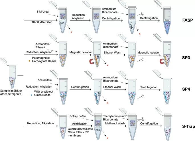
Figure 3. Detergent-removal workflows for bottom-up membrane proteomics. FASP, SP3, SP4, and S-Trap use different filter-, bead-, precipitation-, and trap-based strategies before LC-MS/MS. Reproduced from Figure 1 in Brough et al. (2024), Biochemical Society Transactions, under CC BY 4.0.
Transmembrane regions remain difficult to digest and detect because they often yield long, hydrophobic peptides. Trypsin remains the default enzyme for bottom-up proteomics, while Lys-C is commonly paired with trypsin because it tolerates strongly denaturing conditions such as high urea concentrations. When tryptic coverage of hydrophobic regions is poor, a complementary protease can generate alternative peptides rather than repeating the same digestion; chymotrypsin is one option because it cleaves after several aromatic or hydrophobic residues. Multi-enzyme workflows can improve sequence coverage, but they also increase peptide complexity and database-search space. They are therefore most useful when additional coverage of membrane-spanning regions or specific membrane targets is a defined objective (Jiang et al., 2024; Miller & Smith, 2023).
After digestion, peptides generally require desalting and removal of residual contaminants before LC-MS/MS. This is especially important after detergent- or lipid-rich extraction, because carryover can impair chromatography or ionization. Offline peptide fractionation can increase analytical depth, but it also adds handling, instrument time, and opportunities for peptide loss or batch variation. Additional cleanup or fractionation is most useful when it addresses a defined interference or depth requirement rather than being added routinely (Jiang et al., 2024).
6. LC-MS/MS Identification and Membrane-Specific Coverage Assessment
Once peptides are MS-ready, acquisition mode affects how consistently proteins are quantified across samples. Data-dependent acquisition (DDA) selects individual precursor ions for fragmentation, whereas data-independent acquisition (DIA) fragments broader m/z windows more systematically, which can improve quantitative completeness and reduce missing values in comparative datasets. This advantage does not compensate for upstream losses: proteins discarded during extraction or peptides lost during cleanup cannot be recovered at the acquisition stage. For membrane proteomics, the key constraint remains whether hydrophobic peptides survive sample preparation in a form that can be chromatographed, ionized, fragmented, and confidently assigned (Jiang et al., 2024).
Total protein IDs provide only a partial view of protocol performance. Membrane-focused evaluation should consider the number and proportion of annotated or integral membrane proteins, recovery of difficult classes such as multi-pass proteins or predefined target families, and quantitative completeness and reproducibility. Transmembrane-helix distribution, hydrophobic-peptide representation, and sequence coverage can show whether higher protein counts reflect deeper sampling of difficult membrane regions rather than simply more soluble proteins. In mouse liver, Antony et al. (2026) compared four solid-phase workflows with three membrane-mimetic approaches and found that methods producing more total protein identifications did not necessarily provide the strongest membrane-protein representation. Global proteome depth and membrane-specific coverage therefore need to be evaluated separately.
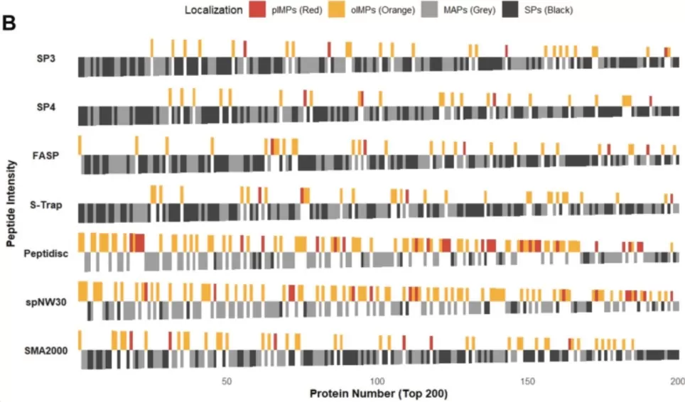
Figure 4. Membrane-protein representation across sample-preparation workflows. The top 200 proteins ranked by MaxLFQ intensity are classified as plasma-membrane integral proteins (pIMPs), organellar integral proteins (oIMPs), membrane-associated proteins (MAPs), or soluble proteins (SPs). Cropped from Figure 3B in Antony et al. (2026), Molecular & Cellular Proteomics, under CC BY 4.0; cropped for layout only.
Protocol comparisons are easier to interpret when biological input and downstream processing are held constant. A gain in membrane-protein coverage still needs to be checked against missing values, quantitative variability, and replicate reproducibility. Non-detection also requires caution: a biologically present protein may disappear from the reported dataset because it was lost during extraction, digestion, cleanup, or peptide detection. Membrane-specific coverage and routine quantitative quality-control measures should therefore be considered together.
7. Common Failure Modes and Troubleshooting in Membrane Protein Proteomics
Troubleshooting should start by locating the earliest likely point of protein or peptide loss in the workflow. If extraction, digestion, or cleanup is limiting recovery, changing acquisition settings alone is unlikely to solve the underlying problem.
Table 2. Troubleshooting Membrane Protein Proteomics: Common Problems, Causes, and Solutions
| Problem | Possible cause | Strategy to consider |
|---|---|---|
| Good total protein yield but few membrane proteins | Soluble proteins dominate; insufficient enrichment | Consider membrane/subcellular enrichment or complementary fractionation |
| Large insoluble pellet after lysis | Incomplete solubilization or heterogeneous insoluble material | Evaluate pellet composition; use stronger or complementary extraction only when aligned with the target |
| Protein pellet is difficult to redissolve | Over-precipitation or over-drying | Optimize precipitation conditions, drying time, and resolubilization chemistry |
| Poor digestion or few membrane-derived peptides | Residual detergent/chaotrope; limited protease accessibility | Improve cleanup or buffer exchange and review digestion conditions |
| LC-MS signal suppression | Residual detergents, lipids, or salts | Strengthen cleanup/desalting and review extraction-to-MS compatibility |
| High between-sample variability | Inconsistent fractionation/extraction or low recovery | Standardize processing order, timing, input amount, and QC checkpoints |
| Many total proteins but poor multi-pass membrane coverage | Extraction or peptide-detection bias | Use membrane-specific enrichment and evaluate transmembrane-protein and peptide coverage rather than total IDs alone |
8. Membrane and Insoluble Protein Extraction FAQs
8.1 Which Detergent Is Best for Membrane Protein Extraction Before LC-MS/MS?
There is no universal best detergent. The appropriate choice depends on the target protein class, sample matrix, whether native structure or interactions must be retained, and how the detergent will be removed or made compatible with digestion and LC-MS/MS. The detergent and the planned cleanup and digestion steps should be selected as a single workflow.
8.2 Should the Insoluble Pellet Be Discarded During Proteomics Sample Preparation?
Not automatically. The pellet may contain membrane proteins and other difficult targets, but it can also contain cytoskeletal material, aggregates, nucleic-acid-associated components, and other insoluble material. Sequential extraction can be useful when those proteins are relevant, but a pellet should not be labeled a membrane fraction without an enrichment rationale.
8.3 When Is Lipidomics Useful Alongside Membrane Proteomics?
Lipidomics is most informative when the question extends beyond protein abundance to membrane composition or lipid remodeling, such as changes in phospholipids, sphingolipids, sterols, or lipid-signaling species. Proteomics can reveal changes in membrane-associated proteins and pathways, while lipidomics captures remodeling of the lipid environment in which those proteins function. The combination is most useful when lipid remodeling is part of the biological mechanism rather than as a routine add-on to every membrane-protein study.
DIA Quantitative Proteomics and Lipidomics for Membrane-Focused Studies
When the study goal is to compare protein abundance across groups, MetwareBio’s DIA Quantitative Proteomics service uses LC-MS/MS-based data-independent acquisition for label-free protein identification and quantification, followed by differential-protein analysis, quality-control assessment, and downstream biological interpretation. In the context of membrane-focused studies, DIA is most useful after sample preparation has already preserved the difficult proteins and peptides that need to be compared across samples.
If the biological hypothesis also includes membrane lipid remodeling, MetwareBio’s Quantitative Lipidomics service can add targeted UPLC-MS/MS-based semi-quantitative profiling of 4,000+ lipid species across 51 classes, including glycerophospholipids, sphingolipids, and sterol lipids. The two assays answer different questions: proteomics measures changes in proteins and pathways, whereas lipidomics measures changes in the membrane lipid environment. A combined design is therefore most informative when both layers are part of the mechanism, not as a routine add-on to every membrane-protein project. MetwareBio can support DIA quantitative proteomics alone or a combined proteomics-lipidomics study according to the biological question and sample type.
Contact UsRead More: Membrane Protein Extraction and Sample Preparation for Proteomics
These articles complement the current guide by covering sample-type-specific protein extraction, digestion and desalting strategies, acquisition-mode selection, and high-throughput sample preparation for membrane-focused and other challenging proteomics studies.
Section 2 explains that sample matrix determines which preparation problem dominates. This guide details extraction workflows for cells, animal and plant tissues, and body fluids, including high-abundance-protein depletion strategies that shape downstream membrane-protein recovery.
Section 5 emphasizes converting extracted proteins into MS-compatible peptides. This article covers reduction and alkylation, trypsin and Lys-C digestion choices, and C18 desalting steps that directly support reliable LC-MS/MS analysis.
Complement the digestion guidance in Section 5 by comparing in-gel and in-solution digestion approaches, including how gel structure affects digestibility and which workflow best fits LC-MS/MS-based identification.
Section 6 discusses how acquisition mode affects quantitative completeness. This article compares DIA and DDA strategies to help you choose the approach best matched to your membrane-proteome coverage goals.
Extend the sample-preparation discussion with a high-throughput perspective, covering scalable extraction, digestion, and cleanup workflows that maintain consistency when membrane-protein studies expand to larger cohorts.
This article focuses on bottom-up identification; this guide explains how top-down proteomics differs in workflow, coverage, and application, helping you position membrane-protein analysis within the broader proteomics landscape.
References
- Antony, F., Bhattacharya, A., Aoki, H., Jandu, R. S., Abdualkader, A. M., Al Batran, R., Babu, M., & Duong van Hoa, F. (2026). Comparative evaluation of solid-phase and membrane mimetic strategies in membrane proteome coverage and disease-state analysis. Molecular & Cellular Proteomics, 25(2), 101496. https://doi.org/10.1016/j.mcpro.2025.101496
- Behnke, J.-S., & Urner, L. H. (2023). Emergence of mass spectrometry detergents for membrane proteomics. Analytical and Bioanalytical Chemistry, 415, 3897-3909. https://doi.org/10.1007/s00216-023-04584-z
- Behnke, J.-S., Hentschel, A., Wolf, M., Wycisk, V., Sickmann, A., Heyer, R. S., & Urner, L. H. (2025). Detergent screening with hybrid detergents increases observable number of protein identities in bottom-up proteomics. Proteomics, 25(15), 6-12. https://doi.org/10.1002/pmic.70003
- Brough, Z., Zhao, Z., & Duong van Hoa, F. (2024). From bottom-up to cell surface proteomics: Detergents or no detergents, that is the question. Biochemical Society Transactions, 52(3), 1253-1263. https://doi.org/10.1042/BST20231020
- Duong, V.-A., & Lee, H. (2023). Bottom-up proteomics: Advancements in sample preparation. International Journal of Molecular Sciences, 24(6), 5350. https://doi.org/10.3390/ijms24065350
- Fang, F., Zhao, Q., Li, X., Liang, Z., Zhang, L., & Zhang, Y. (2016). Dissolving capability difference based sequential extraction: A versatile tool for in-depth membrane proteome analysis. Analytica Chimica Acta, 945, 39-46. https://doi.org/10.1016/j.aca.2016.09.032
- Jiang, Y., Rex, D. A. B., Schuster, D., Neely, B. A., Rosano, G. L., Volkmar, N., Momenzadeh, A., Peters-Clarke, T. M., Egbert, S. B., Kreimer, S., Doud, E. H., Crook, O. M., Yadav, A. K., Vanuopadath, M., Hegeman, A. D., Mayta, M. L., Duboff, A. G., Riley, N. M., Moritz, R. L., & Meyer, J. G. (2024). Comprehensive overview of bottom-up proteomics using mass spectrometry. ACS Measurement Science Au, 4(4), 338-417. https://doi.org/10.1021/acsmeasuresciau.3c00068
- Kongpracha, P., Wiriyasermkul, P., Isozumi, N., Moriyama, S., Kanai, Y., & Nagamori, S. (2022). Simple but efficacious enrichment of integral membrane proteins and their interactions for in-depth membrane proteomics. Molecular & Cellular Proteomics, 21(5), 100206. https://doi.org/10.1016/j.mcpro.2022.100206
- Masuda, T., Ito, S., & Ohtsuki, S. (2021). Advances in sample preparation for membrane proteome quantification. Drug Discovery Today: Technologies, 39, 23-29. https://doi.org/10.1016/j.ddtec.2021.06.005
- Miller, R. M., & Smith, L. M. (2023). Overview and considerations in bottom-up proteomics. Analyst, 148(3), 475-486. https://doi.org/10.1039/D2AN01246D
- Varnavides, G., Madern, M., Anrather, D., Hartl, N., Reiter, W., & Hartl, M. (2022). In search of a universal method: A comparative survey of bottom-up proteomics sample preparation methods. Journal of Proteome Research, 21(10), 2397-2411. https://doi.org/10.1021/acs.jproteome.2c00265