Protein glycosylation is one of the most biologically pervasive post-translational modifications. It shapes protein folding and stability, modulates receptor–ligand recognition, influences immune signaling, and can remodel cell–cell communication in development and disease [1]. In mass spectrometry (MS)-based glycoproteomics, however, glycosylation is also a major analytical challenge. Glycopeptides are often low in abundance, structurally heterogeneous, and easily suppressed by high-abundance non-modified peptides during LC–MS/MS. As a result, direct injection of a complex digest typically yields poor coverage of glycosylation sites and glycoform diversity.
This is why glycopeptide enrichment is widely regarded as a decisive pre-analytical step for confident glycoproteomics. The goal is not merely to “concentrate” glycopeptides, but to increase selectivity, reduce matrix interference, and deliver spectra rich enough to support intact glycopeptide identification (peptide sequence + glycosylation site + glycan composition) or, when desired, site-focused workflows after deglycosylation. Among many approaches, lectin affinity chromatography and hydrophilic interaction chromatography (HILIC) have become two mainstream strategies because they are conceptually robust, experimentally accessible, and broadly compatible with modern LC–MS platforms [2,3].
In this guide, we expand and optimize a practical comparison of these two methods—covering principles, strengths and limitations, ideal applications, and how MS/MS fragmentation strategies can validate and complement enrichment performance.
1. Challenges in LC–MS/MS Glycopeptide Analysis
Before comparing enrichment tools, it helps to clarify what “complexity” means in glycoproteomics. A single glycosylation site can carry many glycoforms (microheterogeneity), and a protein can contain multiple glycosylation sites (macroheterogeneity). This diversity is biologically meaningful—yet analytically it spreads signal across many related species, each potentially appearing at low intensity. Glycans are also chemically distinct from peptides: they are highly polar and can fragment readily, sometimes dominating MS/MS spectra and obscuring peptide backbone ions.
Enrichment is therefore designed to improve the probability that glycopeptides are selected for fragmentation and that the resulting spectra are interpretable. Practically, a “good” enrichment should deliver high recovery, strong selectivity, and minimal bias against specific glycoforms or peptide chemistries. The catch is that no single method perfectly satisfies all three—so the best choice depends on whether your goal is targeted confirmation of known glycan motifs, or discovery-driven, unbiased profiling of a complex glycoproteome.
2. Lectin Affinity Chromatography for Glycopeptide Enrichment
2.1 Principle and Workflow of Lectin-Based Enrichment
Lectins are glycan-binding proteins that recognize specific carbohydrate motifs through reversible, noncovalent interactions. They occur in plants, animals, and microorganisms and comprise diverse structural families with different glycan-binding specificities. For glycopeptide enrichment, the binding specificity of the selected lectin is generally more important than its biological origin [2,3].
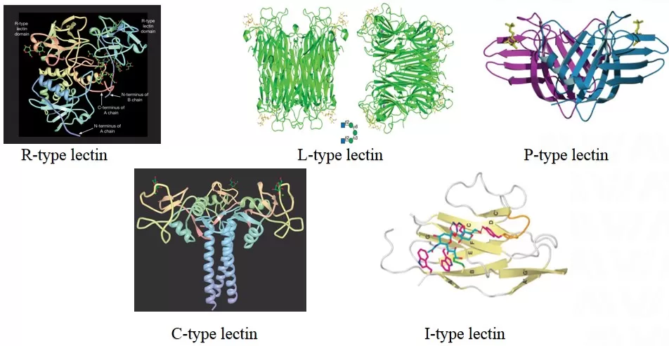
Figure 1. Common lectins: R-type, L-type; P-type, C-type, I-type [2].
In lectin affinity chromatography, one or more lectins are immobilized on a solid support. When a peptide mixture passes through, glycopeptides carrying compatible glycan motifs bind to the lectin, while most non-glycosylated peptides flow through. After washing, bound glycopeptides are eluted by disrupting lectin–glycan binding (often via competitive sugars, altered ionic strength, or pH adjustments) [4,5].
This mechanism gives lectin enrichment a distinctive advantage: biological selectivity. For example, ConA is commonly associated with high-mannose and hybrid N-glycans, while WGA can recognize N-acetylglucosamine and sialic acid-related motifs. Because lectins “see” glycan features rather than generic hydrophilicity, lectin workflows can be tuned to answer motif-specific questions, such as whether a disease state shows increased high-mannose occupancy or altered sialylation patterns.
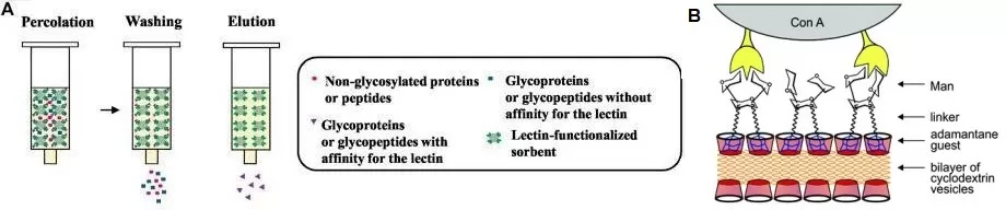
Figure 2. A: General workflow of lectin solid-phase extraction. First, precondition the lectin sorbent; then load (permeate) the sample onto the sorbent and wash away components that do not react with the lectin; finally, elute the target glycoproteins or glycopeptides from the sorbent by disrupting the lectin–carbohydrate binding interaction [3]. B: Schematic of ConA binding to high-mannose type [4]
2.2 Key Advantages: Glycan Motif Selectivity
Lectin affinity chromatography is especially powerful when you already have a hypothesis about glycan features. In biomarker-oriented studies, where researchers focus on specific glycoforms known to associate with inflammation, cancer, or immune activation, lectin enrichment can improve interpretability because it reduces the chemical diversity entering the MS. This often translates into cleaner spectra and lower background noise. Modern lectin-based affinity sorbents—commercial or custom—have also matured in terms of material design and workflow standardization, supporting both glycoprotein and glycopeptide capture depending on where lectins are placed in the pipeline (protein-level vs peptide-level enrichment).
2.3 Key Limitations: Coverage Bias and Specificity Constraints
The same selectivity that makes lectins attractive can also impose bias. Lectins cannot enrich glycoforms they do not recognize, and many lectins have overlapping—but not identical—specificities. In discovery-mode glycoproteomics, this can lead to incomplete coverage of the glycoproteome, especially if novel or unexpected glycan motifs are important to the biology. Additionally, lectin binding can be influenced by steric context and sample composition, which means performance may vary across matrices such as plasma, tissue lysate, or secretome. For broad profiling, lectins are often used in multi-lectin combinations to expand glycoform coverage, but that increases experimental complexity and can complicate reproducibility if not carefully standardized.
3. Hydrophilic Interaction Chromatography (HILIC) Based Glycopeptide Enrichment
3.1 Principle and Workflow of HILIC-Based Enrichment
HILIC relies on the high hydrophilicity of glycans. Most HILIC stationary phases are polar (often silica-based) and support the formation of a water-enriched layer on the surface under high-organic mobile phase conditions. Analytes partition between the bulk organic-rich mobile phase and this hydrophilic layer. Glycopeptides, enriched in hydroxyl-bearing glycans, preferentially interact with the hydrophilic environment and are retained longer than many non-glycosylated peptides.
In zwitterionic HILIC (commonly discussed in glycopeptide work), retention may involve a combination of hydrophilic partitioning, hydrogen bonding, and weak electrostatic interactions. This blended interaction profile helps explain why HILIC can provide broad glycopeptide capture across different glycan types and degrees of sialylation.
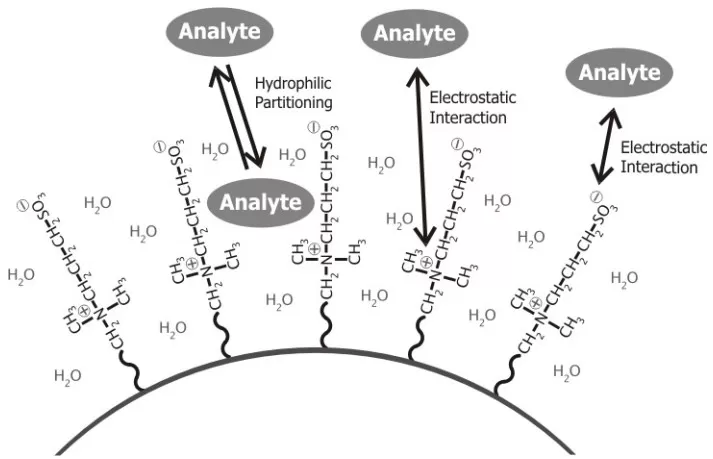
Figure 3. HILIC retention mechanism on a zwitterionic stationary phase via hydrophilic partitioning and electrostatic interactions
(A practical guide to hydrophilic interaction chromatography (HILIC): Wen Jiang, 2005)
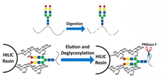
Figure 4. Enrichment process of HILIC technology [6]
3.2 Key Advantages: Broad-Spectrum Glycopeptide Coverage
HILIC is widely adopted for global (discovery-driven) glycoproteomics because it is comparatively broad-spectrum. Unlike lectins, it does not require prior assumptions about glycan motifs. This makes it suitable for screening unknown glycoforms, comparing disease vs control cohorts, and mapping glycosylation landscapes in tissues, biofluids, and cell models [6][7]. Another practical advantage is that HILIC can preserve glycopeptides intact, making it compatible with workflows that aim to identify both glycosites and glycan compositions without obligatory glycan removal.
From an operational standpoint, HILIC is also amenable to high-throughput and automation. Many labs implement HILIC-based enrichment in SPE cartridges or StageTips, and it integrates naturally with LC–MS/MS pipelines designed for scale.
3.3 Key Limitations: Method Sensitivity and Co-Enrichment
HILIC enrichment is powerful, but it is sensitive to conditions. The organic solvent proportion, buffer composition, and sample cleanup can strongly affect both selectivity and recovery. Highly hydrophobic glycopeptides may show weaker retention, while extremely polar contaminants can co-enrich, especially if desalting and detergent removal are incomplete. As HILIC formats diversify, method development becomes part of success: a “one-size-fits-all” recipe is rarely optimal for every matrix, instrument, or glycoproteome depth target.
4. Lectin Affinity vs HILIC: Key Differences for Glycoproteomics
Below is a refined comparison to help match strategy to research intent. The key is to treat enrichment as a decision tool: it should align with your biological question, sample type, and downstream MS workflow.
Table 1. Comparison of Lectin Affinity Chromatography and HILIC for Glycopeptide Enrichment [3–8]
| Dimension | Lectin affinity chromatography | Hydrophilic interaction chromatography (HILIC) |
|---|---|---|
| Core principle | Specific recognition of glycan motifs through noncovalent lectin-glycan interactions | Based on glycan hydrophilicity; glycopeptides are retained on a hydrophilic stationary phase under high-organic conditions |
| Recognition mechanism | Lectin-dependent glycan motif recognition (e.g., ConA binds selected mannose-rich glycans; WGA recognizes GlcNAc-containing motifs) | Hydrophilic partitioning, with hydrogen bonding and electrostatic interactions depending on the stationary phase |
| Glycoform coverage | Enriches glycopeptides carrying glycan motifs recognized by the selected lectin(s); coverage is lectin-dependent | Can enrich many N- and O-linked glycopeptides, but recovery varies with glycan and peptide properties; coverage is not universal |
| Structural integrity | Can preserve intact glycopeptides during capture and elution; deglycosylation is optional and depends on the downstream analysis | Can preserve intact glycopeptides; deglycosylation may be added for glycosylation-site-focused workflows |
| Enrichment efficiency | Selectivity and recovery depend on lectin specificity, binding affinity, and sample conditions | Recovery and selectivity vary with glycopeptide properties and enrichment conditions; nonglycosylated peptides may co-enrich |
| Throughput and automation | Can be implemented in single- or multi-lectin workflows; throughput depends on the enrichment format and automation | Compatible with SPE, StageTip, and automated workflows; throughput depends on the enrichment format |
| Sample compatibility | Applicable to complex samples, but performance depends on glycan accessibility, sample composition, and preprocessing | Suitable for diverse sample matrices, including serum, tissues, and cell lysates, with appropriate sample preparation |
| Suitable research type | Hypothesis-driven analysis of selected glycan motifs and glycoform-related changes | Discovery-oriented glycopeptide profiling and broad comparative glycoproteomics |
| Main advantages | Motif-selective enrichment; reduced sample complexity; useful for hypothesis-driven glycan-feature studies | Broad glycopeptide capture without predefined glycan motifs; compatible with intact glycopeptide analysis and scalable workflows |
| Main limitations | Limited to glycan motifs recognized by the selected lectin(s); binding may vary with steric accessibility and sample composition | Recovery depends on glycan size, peptide properties, stationary phase, and loading conditions; hydrophilic nonglycosylated peptides may co-enrich |
| Downstream MS compatibility | Compatible with intact glycopeptide analysis or deglycosylation-based site mapping, depending on the workflow | Compatible with intact glycopeptide analysis or deglycosylation-based site mapping, depending on the workflow |
5. MS/MS Fragmentation for Glycopeptide Identification After Enrichment
Even with excellent enrichment, identification quality depends on fragmentation. Glycopeptides fragment differently from standard peptides: glycans can dominate fragmentation channels, while peptide backbone ions are essential for confident sequence and site assignment. For this reason, glycoproteomics often benefits from combining dissociation modes, or applying hybrid schemes to extract both glycan and peptide information from a single precursor [9-11].
Common dissociation modes include collision-induced dissociation (CID), higher-energy collisional dissociation (HCD), stepped collision energy HCD (sceHCD), electron transfer dissociation (ETD), and electron transfer/higher-energy collisional dissociation (EThcD)—a hybrid that aims to recover complementary peptide and glycan fragments in one spectrum. Recent comparative studies suggest that hybrid or sequential strategies can improve performance for complex intact glycopeptides, especially when both glycosite localization and glycan composition are needed.
Table 2 Comparison of four major dissociation modes
| Mode | Core mechanism | Glycan fate | Information obtained | Best application scenario |
|---|---|---|---|---|
| CID | Low-energy collision | Preferential glycan cleavage | Glycan fragments (oxonium ions), limited backbone | Glycan-feature screening, quick confirmation |
| HCD | High-energy collision | Extensive cleavage | Glycan fragments + partial backbone | Glycan structural clues + partial peptide evidence |
| ETD | Electron transfer | Glycan largely preserved | Strong peptide backbone, glycosite localization | Site-centric mapping, PTM-friendly workflows |
| EThcD | ETD + HCD combination | Controllable cleavage | More complete intact glycopeptide characterization | Deep profiling, microheterogeneity resolution |
In practical terms, the enrichment method and fragmentation strategy should be considered together. For example, HILIC often yields diverse intact glycopeptides that benefit from EThcD or optimized HCD/sceHCD workflows, while lectin enrichment may reduce glycan diversity and simplify interpretation even with conventional HCD settings.
6. How to Choose a Glycopeptide Enrichment Method
If your primary goal is discovery—to map glycosylation sites broadly, compare conditions, or identify unexpected glycoform shifts—HILIC is usually the most straightforward starting point. It tends to capture a wide range of glycopeptides across complex matrices and supports intact glycopeptide analysis workflows commonly used in modern glycoproteomics [6,7]. When your results reveal a specific glycan feature that appears biologically important (for example, shifts in high-mannose occupancy or altered sialylation signatures), lectin enrichment can then serve as a targeted follow-up to increase selectivity and improve validation confidence.
If your study is already motif-focused—such as interrogating a known glycan hallmark or enriching a glycan subtype relevant to your hypothesis—lectin affinity chromatography often delivers higher interpretability and cleaner MS backgrounds. In those cases, selecting appropriate lectins (and, when necessary, employing multi-lectin strategies) becomes the core experimental design step rather than a minor technical choice.
Finally, regardless of enrichment, do not underestimate sample prep fundamentals. Glycopeptide enrichment is highly sensitive to contaminants that disrupt binding or suppress ionization. In practice, careful desalting, detergent removal, and consistent loading amounts often explain more performance variability than the choice between lectin and HILIC itself.
Plan Your N-Glycosylation Proteomics Study with MetwareBio
Planning an N-glycosylation proteomics study? MetwareBio provides HILIC-based N-glycopeptide enrichment followed by enzymatic deglycosylation and 4D label-free LC-MS/MS for N-glycosylation site identification and comparative quantification. Our team can help evaluate sample requirements, experimental design, and site-level analysis options. If your project requires intact glycopeptide or glycoform composition data, discuss these analytical requirements before selecting a workflow.
Explore N-Glycosylation Proteomics ServicesRead More: Glycoproteomics and Glycopeptide Analysis
These articles expand on glycopeptide enrichment, fragmentation strategies, and the broader glycoproteomics workflows that turn enriched glycopeptides into confident biological conclusions.
A broader overview of LC–MS/MS-based glycoproteomics: intact glycopeptide workflows, N- and O-glycosylation mapping, and how site-specific biomarkers are used across diseases.
Covers enrichment and ETD/EThcD fragmentation for a labile intracellular glycosylation—a concrete example of how enrichment choice and dissociation strategy must be designed together.
Compares the structural logic, biosynthesis, and analytical strategies for N- and O-glycans—the glycan-biology background behind lectin and HILIC selectivity.
Shows how glycosylation patterns serve as disease biomarkers in cancer and immune disorders, motivating the targeted and discovery enrichment workflows compared in this article.
Positions glycopeptide enrichment within the full bottom-up LC–MS/MS pipeline, from digestion and cleanup to DDA, DIA, and PRM acquisition.
Details what happens after acquisition: database search, FDR control, and quantification—the computational side of interpreting enriched glycopeptide datasets.
References
- Alter, Galit et al. “Antibody glycosylation in inflammation, disease and vaccination.” Seminars in immunology vol. 39 (2018): 102-110. https://doi.org/10.1016/j.smim.2018.05.003
- Varki, A., Cummings, R. D., Esko, J. D., et al. (Eds.). (2022). Glycan-binding proteins. In Essentials of Glycobiology (4th ed.). Cold Spring Harbor Laboratory Press.
- Goumenou, Anastasia et al. “Recent Advances in Lectin-Based Affinity Sorbents for Protein Glycosylation Studies.” Frontiers in molecular biosciences vol. 8 746822. 29 Oct. 2021, https://doi.org/10.3389/fmolb.2021.746822.
- Bojarová, P, and V Křen. “Sugared biomaterial binding lectins: achievements and perspectives.” Biomaterials science vol. 4,8 (2016): 1142-60. https://doi.org/10.1039/c6bm00088f.
- Ruiz-May, Eliel et al. “A comparative study of lectin affinity based plant N-glycoproteome profiling using tomato fruit as a model.” Molecular & cellular proteomics : MCP vol. 13,2 (2014): 566-79. https://doi.org/10.1074/mcp.M113.028969.
- Zhu, Jun et al. “Comprehensive mapping of protein N-glycosylation in human liver by combining hydrophilic interaction chromatography and hydrazide chemistry.” Journal of proteome research vol. 13,3 (2014): 1713-21. https://doi.org/10.1021/pr401200h.
- Chen, Zhengwei et al. “In-depth Site-specific Analysis of N-glycoproteome in Human Cerebrospinal Fluid and Glycosylation Landscape Changes in Alzheimer's Disease.” Molecular & cellular proteomics : MCP vol. 20 (2021): 100081. https://doi.org/10.1016/j.mcpro.2021.100081.
- Zhu, Fangli et al. “Integrated Proteomic and N-Glycoproteomic Analyses of Chicken Egg during Embryonic Development.” Journal of agricultural and food chemistry vol. 67,42 (2019): 11675-11683. https://doi.org/10.1021/acs.jafc.9b05133.
- Wang, Yue, and Zhixin Tian. “New Energy Setup Strategy for Intact N-Glycopeptides Characterization Using Higher-Energy Collisional Dissociation.” Journal of the American Society for Mass Spectrometry vol. 31,3 (2020): 651-657. https://doi.org/10.1021/jasms.9b00089
- Riley, Nicholas M et al. “Optimal Dissociation Methods Differ for N- and O-Glycopeptides.” Journal of proteome research vol. 19,8 (2020): 3286-3301. https://doi.org/10.1021/acs.jproteome.0c00218.
- Zeng, Wenjuan et al. “Comparative N-Glycoproteomics Analysis of Clinical Samples Via Different Mass Spectrometry Dissociation Methods.” Frontiers in chemistry vol. 10 839470. 24 Feb. 2022, https://doi.org/10.3389/fchem.2022.839470.