Plant lipids shape cellular membranes, store carbon and energy, and participate in signaling throughout growth and environmental adaptation. Their biological effects depend on the composition and distribution of individual lipid species, which can change even when total lipid content remains similar. Understanding these changes is important for investigating plant development, stress tolerance, seed oil composition, and crop quality. Plant lipidomics provides a systematic way to examine this molecular diversity, but its usefulness depends on matching analytical coverage and data interpretation to the biological question. This article explains the major plant lipid groups, the principles of lipidomics analysis, and representative applications in plant research. It helps researchers understand what lipid profiling can reveal, recognize the factors that influence results, and plan studies that connect lipid changes with meaningful biological questions.
1. What Is Plant Lipidomics?
Plant lipidomics is the systematic study of lipid composition, abundance, and remodeling in plant cells, tissues, and organs. It characterizes lipids at the class and molecular-species levels, providing information about how plants organize membranes, store resources, and respond to developmental or environmental signals. Its value lies in connecting changes in lipid chemistry with the biological processes occurring in the sampled material.
This molecular detail complements conventional measurements of total oil and fatty acid composition. Total oil content indicates the size of a lipid pool, while total fatty acid analysis describes its constituent acyl chains. Intact-lipid profiling adds information about how those chains are distributed among different lipid classes and molecular species. For example, changes in an unsaturated fatty acid within chloroplast membrane lipids and storage triacylglycerols can reflect different aspects of lipid metabolism. Measuring these pools separately helps distinguish changes in membrane composition from changes in lipid storage and turnover (Bengtsson et al., 2021).
Plant lipidomics also complements conventional plant metabolomics through workflows specifically optimized for lipid analysis. Lipid-oriented extraction, chromatographic separation, mass spectrometric acquisition, and dedicated annotation strategies generally provide broader lipid coverage and more detailed molecular characterization than conventional metabolomics workflows, enabling more comprehensive analysis of plant lipid composition and remodeling (Herrfurth et al., 2021).
These capabilities support comparisons among genotypes, tissues, developmental stages, and treatments. A lipid profile provides a molecular phenotype that can be examined alongside physiological traits and gene or protein expression. Coordinated changes across lipid classes can suggest how carbon allocation, membrane composition, or lipid turnover differs between conditions, helping researchers prioritize processes for functional investigation. Developmental lipid maps illustrate how this information can establish a biological reference for interpreting plant lipid variation (Kehelpannala et al., 2021).
2. Plant Lipid Classes and Biological Functions
Plant lipids span several major chemical categories, including fatty acyls, glycerolipids, glycerophospholipids, sphingolipids, sterol lipids, and prenol lipids (Liebisch et al., 2020). Their structural diversity supports a wide range of biological functions: membrane lipids provide the physical framework for cellular compartments, storage lipids supply carbon and energy, and other lipid molecules participate in signaling, surface protection, photosynthesis, and antioxidant defense. Table 1 summarizes these major categories, representative plant lipids, and their principal biological roles.
| Lipid category | Representative plant lipids | Principal biological roles |
|---|---|---|
| Fatty acyls (FA) | Free fatty acids, oxylipins, fatty alcohols, and wax esters | Supply acyl building blocks and participate in signaling or surface protection, depending on the subclass. |
| Glycerolipids (GL) | Mono-, di-, and triacylglycerols (MAG, DAG, TAG); monogalactosyldiacylglycerol (MGDG), digalactosyldiacylglycerol (DGDG), and sulfoquinovosyldiacylglycerol (SQDG). | Connect lipid storage with membrane metabolism and include major chloroplast glycolipids. |
| Glycerophospholipids (GP) | Phosphatidylcholine (PC), phosphatidylethanolamine (PE), phosphatidylglycerol (PG), and phosphatidic acid (PA) | Form cellular membranes; selected classes also participate in lipid synthesis and signaling. |
| Sphingolipids (SP) | Ceramides, glucosylceramides, and glycosylinositolphosphoceramides (GIPCs) | Contribute to membrane organization and processes associated with cellular signaling and stress responses. |
| Sterol lipids (ST) | Phytosterols, sterol esters, and sterol glycosides | Influence membrane organization and physical properties, with conjugated forms contributing to sterol homeostasis. |
| Prenol lipids (PR) | Carotenoids, tocopherols, plastoquinone, and ubiquinone | Participate in light harvesting, photoprotection, antioxidant defense, and electron transfer, depending on the compound. |
The relative importance of these groups depends on tissue function. Photosynthetic tissues contain substantial chloroplast membrane systems, making their glycolipid composition particularly informative, while seeds often emphasize storage lipid accumulation. Within membranes, interactions between classes also matter. Biophysical experiments by Mamode Cassim and colleagues (2021), for example, showed that plant GIPCs interact with phytosterols and influence model membrane properties. Among acyl-containing lipids, fatty-acyl chain length and degree of unsaturation add further molecular diversity. An unchanged lipid-class total can therefore conceal shifts among molecular species with different physical or metabolic properties. Consistent lipid classification and nomenclature help preserve these distinctions and connect analytical results with specific biological questions.
3. Plant Lipidomics Analysis: Key Considerations
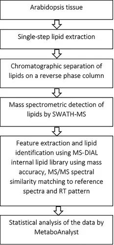
Plant lipidomics connects sampling and preservation with lipid extraction, liquid chromatography–tandem mass spectrometry (LC-MS/MS) analysis, and biological interpretation. The general lipidomics workflow explains these shared steps; plant studies require particular attention to analytical coverage, sample preservation, and quantitative comparability.
3.1 Lipid Coverage and Assay Selection
Plant lipid coverage needs to reflect the biological process under investigation. Chloroplast studies require appropriate representation of galactolipids, SQDG, and PG, while seed oil research may prioritize TAG molecular species and unusual fatty acids. Investigations of low-abundance signaling lipids or complex sphingolipids can require additional analytical optimization.
A large database alone does not establish that every relevant lipid is measured reliably. Extraction, ionization, acquisition settings, and annotation criteria determine which compounds enter the final dataset. Plant-specific transition lists and reference information can support targeted profiling, but their suitability depends on the lipid structures and classes included. Assessing coverage against the research question is therefore more informative than selecting an assay solely by its total target count (Herrfurth et al., 2021).
3.2 Plant Sampling and Lipid Preservation
Plant sample collection needs to account for organ identity, developmental stage, tissue region, and harvest time. These variables can influence lipid composition independently of the experimental treatment. Consistent sampling is especially important when comparing genotypes with different growth rates or tissues collected across light–dark transitions.
Tissue disruption also exposes lipids to enzymes and conditions that can promote hydrolysis or oxidation. Rapid stabilization and controlled handling help preserve the sampled state, while extraction conditions must limit subsequent degradation. Leaves, roots, and oil-rich seeds differ in water content and matrix composition, so the suitability of a preparation method needs assessment for the material being studied. Lipid extraction methods should therefore be selected for the target lipid classes and plant matrix (Bengtsson et al., 2021).
3.3 Lipid Quantification and Comparability
Quantitative comparisons require a normalization basis that fits the biological question. During dehydration, lipid abundance expressed per unit fresh weight can increase partly because water has been lost. Recording sample mass and, where relevant, moisture content helps distinguish this effect from changes in lipid accumulation.
Cross-tissue comparisons introduce additional uncertainty because different extracts can produce different ionization responses. Appropriate internal standards and quality assessment help evaluate comparability, but cannot automatically eliminate every matrix effect. The reported measurement level also matters: relative signals and semi-quantitative estimates support defined comparisons, while absolute amounts require suitable calibration and validation. Clearly reporting these lipid quantification strategies helps prevent analytical differences from being interpreted as biological changes (Köfeler et al., 2021).
4. Applications of Plant Lipidomics
Plant lipidomics is most informative when molecular lipid changes are linked to a defined biological process. Development, stress responses, and seed oil formation illustrate how this information complements conventional physiological and biochemical measurements.
4.1 Plant Growth and Development
Developmental lipidomics examines how plants allocate lipids as tissues acquire, maintain, or change their functions. Expanding photosynthetic tissues require different membrane systems from storage-rich seeds, and a developing organ can alter its lipid profile as its physiological role changes. Measuring lipid species across a developmental series provides a way to describe these transitions and identify the lipid groups most closely associated with them.
Kehelpannala et al. (2021) analyzed seven Arabidopsis tissues at selected developmental stages and created a publicly accessible lipid map. Alongside distinct tissue inventories, the study documented consumption of TAG reserves during seedling growth, connecting molecular profiling with the mobilization of stored resources. The map provides a reference for identifying lipid pools that change during development and for investigating how storage and membrane metabolism contribute to organ function. Its quantitative developmental comparisons were restricted to the same tissue type because different tissue matrices could influence mass-spectrometric responses.
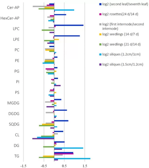
Such developmental information helps distinguish an unusual lipid phenotype from the normal trajectory of the sampled organ. A genotype with delayed development, for example, may differ from a control partly because the tissues are physiologically mismatched. Interpreting lipid changes alongside developmental status and organ function makes candidate metabolic explanations more credible. The resulting hypotheses can guide experiments on membrane formation, storage mobilization, or lipid turnover without treating every developmental association as a direct regulatory mechanism.
4.2 Plant Stress Responses and Defense
Stress lipidomics examines how membrane and storage lipid pools change during environmental challenge or pathogen attack. Differences in acyl composition, lipid classes, and lipid turnover can reveal responses that total lipid measurements conceal. Their interpretation depends on timing and physiological context: a change associated with acclimation can resemble one produced by tissue injury, so lipid profiles gain meaning when considered alongside stress severity and plant performance.
Scholz et al. (2025) investigated Arabidopsis leaves exposed to heat stress or infection by Botrytis cinerea or Pseudomonas syringae, combining lipid analysis with lipid-droplet proteomics. TAG accumulation occurred across the evaluated stresses, accompanied by remodeling of acyl-chain and sterol composition. Changes in lipid-droplet-associated proteins added another view of the response. These findings show why storage lipids and lipid droplets are relevant to stress studies even in vegetative tissues, alongside the membrane lipids more commonly emphasized.
The combined lipid and protein response places lipid droplets within a broader reorganization of cellular metabolism. Changes in TAG composition can be considered together with membrane lipid changes to investigate whether acyl chains are redistributed among pools, while changes in droplet-associated proteins suggest candidate processes involved in storage and turnover. These relationships help researchers investigate how plants manage lipid resources during stress and recovery. Demonstrating that a particular lipid change improves tolerance still requires functional evidence.
4.3 Seed Oil Composition and Crop Quality
Seed lipidomics investigates how fatty acids are incorporated into storage lipids and how molecular composition relates to oil traits. Total oil content describes the size of the storage pool, while fatty acid composition describes its building blocks. Intact-lipid measurements add information about their distribution among TAG, DAG, and membrane lipids, helping locate metabolic constraints that would remain hidden in either bulk measurement alone.
Parchuri et al. (2024) examined how Physaria fendleri accumulates hydroxy fatty acids in seed oil. Their biochemical, genetic, and cell-biological experiments identified a TAG remodeling mechanism involving selective removal of common fatty acids and subsequent incorporation of hydroxy fatty acids. This work shows that final oil composition can be modified after initial TAG synthesis, extending the interpretation of storage lipids beyond their role as metabolic endpoints. It also explains why increasing synthesis of a desired fatty acid may be insufficient if its transfer into storage lipids remains constrained.
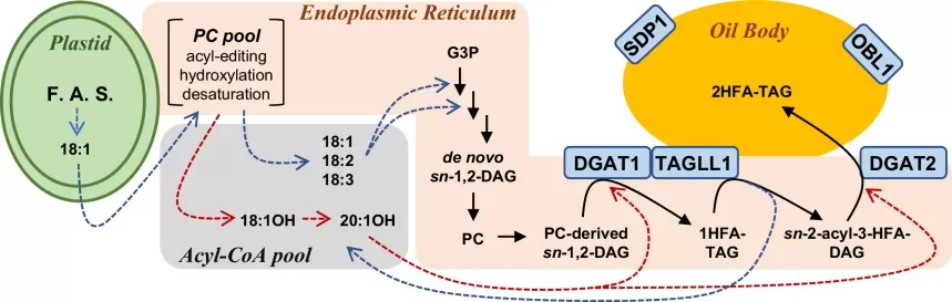
Lipidomics can therefore help evaluate breeding materials or engineered lines by connecting desired oil traits to specific molecular changes. Its relevance also extends to harvested produce. Zhang et al. (2023) combined membrane lipid measurements with physiological and biochemical analyses in cold-stored cucumber, linking fucoidan treatment to altered membrane lipid metabolism and reduced chilling injury. Together, these examples illustrate how lipid analysis can support oil-composition research and postharvest quality studies when integrated with the appropriate phenotype.
5. FAQs About Plant Lipidomics
5.1 Can Dried Plant Samples Be Used for Lipidomics?
Dried plant material can be analyzed when the drying procedure and storage conditions are suitable for the target lipids. Freeze-drying, air-drying, and heat-drying should not be assumed equivalent because oxidation and enzymatic changes may alter the profile. Use consistent preparation across comparison groups, document the treatment, and confirm compatibility with the laboratory before collection.
5.2 Does Plant Lipidomics Cover Cutin, Suberin, and Surface Waxes?
Routine extraction-based lipidomics mainly measures soluble, extractable lipids. Surface waxes may require dedicated extraction and analysis, while cutin and suberin are polymers whose constituent monomers generally require depolymerization before characterization. A broad LC-MS lipid panel therefore does not establish coverage of these materials. Define whether the question concerns membrane lipids, storage lipids, wax composition, or polymer constituents before selecting the workflow (Baales et al., 2021).
5.3 Can Plant Lipidomics Reveal Spatial Lipid Distributions?
Bulk lipidomics measures the composition of the extracted material and loses its original spatial arrangement. Mass spectrometry imaging (MSI) can map lipid-associated signals directly in tissue sections. Wang et al. (2022), for example, visualized heterogeneous lipid distributions in peanut seeds using MALDI-MSI. Such imaging complements molecular profiling, although annotation confidence and quantitative comparability need separate assessment. Explore spatial lipidomics when localization is central to the research question.
Plant Lipidomics Services at MetwareBio
MetwareBio’s plant lipidomics service combines a database of 1,747 plant lipids across six major categories and 31 subclasses with LC-MS/MS analysis and multiple reaction monitoring (MRM)-based semi-quantification. Quality control and downstream data analysis support comparative studies of plant development, stress responses, and crop traits. The lipids detected vary with tissue composition and analytical conditions. Discuss your plant species, sample type, and priority lipid classes with MetwareBio to explore an analysis suited to your research question.
Contact UsRead More: Plant Lipidomics and Lipid Analysis
These articles extend the analytical and biological themes covered here, from plant-specific lipid profiling and extraction chemistry to the statistical and pathway-level interpretation of lipid datasets.
MetwareBio's plant lipidomics service uses a reference database of 1,747 plant lipids across six major categories and 31 subclasses, with LC-MS/MS analysis and MRM-based semi-quantification. Actual coverage varies with sample type and analytical conditions. Explore the service for studies of membrane biology, stress responses, seed oil composition, and crop quality.
A structured walkthrough of lipid classes and their biological roles, followed by the analytical options behind lipid profiling. Useful background for the class-level and molecular-species distinctions that drive plant lipid interpretation.
Covers core lipidomics concepts, sample preparation and extraction, shotgun versus LC-MS strategies, and common pitfalls. A practical starting point if you are planning a first plant lipid profiling experiment.
Compares LC-MS and GC-MS platforms, untargeted and targeted strategies, and the full workflow from sample preparation to data analysis. Helps you judge what a given quantitative lipidomics assay can and cannot report.
A practical comparison of the two dominant extraction chemistries, including recovery by lipid class, phase separation, toxicity, and automation. Directly relevant to the plant matrix and preservation issues raised in the sampling section.
Walks through quality control and preprocessing, univariate and multivariate statistics, and pathway-level interpretation of lipid datasets. Complements the quantification and comparability discussion in this article.
References
- Baales, J., Zeisler-Diehl, V. V., & Schreiber, L. (2021). Analysis of extracellular cell wall lipids: Wax, cutin, and suberin in leaves, roots, fruits, and seeds. Methods in Molecular Biology, 2295, 275–293. https://doi.org/10.1007/978-1-0716-1362-7_15
- Bengtsson, J. D., Wallis, J. G., & Browse, J. (2021). Lipid isolation from plants. Methods in Molecular Biology, 2295, 3–13. https://doi.org/10.1007/978-1-0716-1362-7_1
- Herrfurth, C., Liu, Y.-T., & Feussner, I. (2021). Targeted analysis of the plant lipidome by UPLC-nanoESI-MS/MS. Methods in Molecular Biology, 2295, 135–155. https://doi.org/10.1007/978-1-0716-1362-7_9
- Kehelpannala, C., Rupasinghe, T., Pasha, A., Esteban, E., Hennessy, T., Bradley, D., Ebert, B., Provart, N. J., & Roessner, U. (2021). An Arabidopsis lipid map reveals differences between tissues and dynamic changes throughout development. The Plant Journal, 107(1), 287–302. https://doi.org/10.1111/tpj.15278
- Köfeler, H. C., Ahrends, R., Baker, E. S., Ekroos, K., Han, X., Hoffmann, N., Holčapek, M., Wenk, M. R., & Liebisch, G. (2021). Recommendations for good practice in MS-based lipidomics. Journal of Lipid Research, 62, 100138. https://doi.org/10.1016/j.jlr.2021.100138
- Liebisch, G., Fahy, E., Aoki, J., Dennis, E. A., Durand, T., Ejsing, C. S., Fedorova, M., Feussner, I., Griffiths, W. J., Köfeler, H., Merrill, A. H., Jr., Murphy, R. C., O’Donnell, V. B., Oskolkova, O., Subramaniam, S., Wakelam, M. J. O., & Spener, F. (2020). Update on LIPID MAPS classification, nomenclature, and shorthand notation for MS-derived lipid structures. Journal of Lipid Research, 61(12), 1539–1555. https://doi.org/10.1194/jlr.S120001025
- Mamode Cassim, A., Navon, Y., Gao, Y., Decossas, M., Fouillen, L., Grélard, A., Nagano, M., Lambert, O., Bahammou, D., Van Delft, P., Maneta-Peyret, L., Simon-Plas, F., Heux, L., Jean, B., Fragneto, G., Mortimer, J. C., Deleu, M., Lins, L., & Mongrand, S. (2021). Biophysical analysis of the plant-specific GIPC sphingolipids reveals multiple modes of membrane regulation. Journal of Biological Chemistry, 296, 100602. https://doi.org/10.1016/j.jbc.2021.100602
- Parchuri, P., Bhandari, S., Azeez, A., Chen, G., Johnson, K., Shockey, J., Smertenko, A., & Bates, P. D. (2024). Identification of triacylglycerol remodeling mechanism to synthesize unusual fatty acid containing oils. Nature Communications, 15, 3547. https://doi.org/10.1038/s41467-024-47995-x
- Scholz, P., Doner, N. M., Gutbrod, K., Herrfurth, C., Niemeyer, P. W., Lim, M. S. S., Blersch, K. F., Schmitt, K., Valerius, O., Shanklin, J., Feussner, I., Dörmann, P., Braus, G. H., Mullen, R. T., & Ischebeck, T. (2025). Plasticity of the Arabidopsis leaf lipidome and proteome in response to pathogen infection and heat stress. Plant Physiology, 197(2), kiae274. https://doi.org/10.1093/plphys/kiae274
- Wang, X., Chen, Y., Liu, Y., Ouyang, L., Yao, R., Wang, Z., Kang, Y., Yan, L., Huai, D., Jiang, H., Lei, Y., & Liao, B. (2022). Visualizing the distribution of lipids in peanut seeds by MALDI mass spectrometric imaging. Foods, 11(23), 3888. https://doi.org/10.3390/foods11233888
- Zhang, Y., Lin, D., Yan, R., Xu, Y., Xing, M., Liao, S., Wan, C., Chen, C., Zhu, L., Kai, W., Chen, J., & Gan, Z. (2023). Amelioration of chilling injury by fucoidan in cold-stored cucumber via membrane lipid metabolism regulation. Foods, 12(2), 301. https://doi.org/10.3390/foods12020301