Phosphatidylserine (PS): Structure, Functions, and Detection
Imagine a tiny molecule that acts as a cellular messenger, a structural component, and even a signal for cell death. Phosphatidylserine (PS), this unassuming phospholipid, plays a multifaceted role in cellular biology. In this article, we will explore the structure, functions, and detection methods of PS, shedding light on its significance in various biological processes.
1. What is Phosphatidylserine?
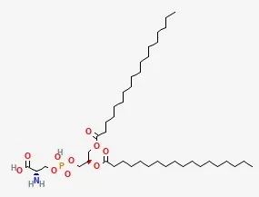
 As we all know, the main component of the cell membrane is the phospholipid bilayer. As a member of this lipid family, phosphatidylserine (PS) plays a key role in the structure of the cell membrane. PS follows a typical structure consisting of a glycerol backbone, two fatty acid chains, and a phosphate group. However, the uniqueness of PS lies in its unique head group, the serine amino acid, which distinguishes it from other phospholipids. This structural feature gives PS unique properties and leads to its asymmetric distribution in the phospholipid bilayer. This asymmetric distribution is essential for various cellular functions, highlighting the importance of PS in maintaining cell health and integrity.
2. Phosphatidylserine Structure
Phosphatidylserine (PS) is an important phospholipid that plays a crucial role in the structure and function of cell membranes. Its unique molecular structure, characterized by a polar head and hydrophobic tail, contributes to its diverse functions and importance in various biological processes.
Molecular structure of phosphatidylserine:
Glycerol skeleton: a three-carbon structure that serves as the skeleton of the entire lipid molecule.
Fatty acid chains: Two hydrophobic fatty acid chains are esterified with the glycerol backbone. The length and saturation of these chains can vary, affecting the fluidity and permeability of the membrane.
Phosphate Group: The phosphate group is attached to the glycerol backbone to form the polar head group.
Amino acid serine: The serine amino acid is linked to a phosphate group, further enhancing the polarity and hydrophilicity of the head group.
Asymmetric distribution in membranes
A distinctive feature of PS is its asymmetric distribution within the lipid bilayer. It is mainly located in the inner leaflet of the plasma membrane. This asymmetry is critical for a variety of cellular processes, including:
-
Apoptosis: PS translocation to the outer leaflet serves as a signal for cell death.
-
Membrane trafficking: PS plays a role in vesicle trafficking and membrane fusion events.
-
Signal transduction: PS can interact with specific proteins to initiate signaling pathways.
3. Function of Phosphatidylserine
1)Membrane Structure and Function
PS is a major component of the inner leaflet of the plasma membrane. Its presence is essential for maintaining membrane fluidity and integrity. It helps to regulate membrane curvature and facilitates the formation of lipid rafts, specialized membrane microdomains that are important for various cellular processes, including signal transduction and receptor clustering.
2) Cell Signaling and Apoptosis
Apoptosis: During apoptosis, PS is translocated from the inner to the outer leaflet of the plasma membrane. This "eat-me" signal is recognized by phagocytes, which engulf and clear apoptotic cells, preventing inflammation.
Signal Transduction: PS can interact with various proteins, such as kinases and phosphatases, to modulate intracellular signaling pathways. It is involved in a range of signaling events, including cell growth, differentiation, and survival.
3) Neuronal Function
Synaptic Transmission: PS plays a critical role in synaptic vesicle fusion, enabling the release of neurotransmitters into the synaptic cleft. This is essential for neuronal communication and synaptic plasticity.
Neurotransmitter Synthesis: PS is involved in the synthesis of certain neurotransmitters, such as acetylcholine.
4) Immune Response
Phagocytosis: The exposure of PS on the cell surface serves as a signal for phagocytosis by immune cells, promoting the clearance of damaged or infected cells.
Immune Cell Activation: PS can modulate the activation and function of immune cells, influencing both innate and adaptive immune responses.
5) Viral Infection
Some viruses, such as HIV, can exploit the exposure of PS on the cell surface to enhance their entry into host cells. This highlights the importance of understanding the role of PS in viral pathogenesis.
4. Differences Between Phosphatidylserine and Other Phospholipids
1) Distribution in Cells:
Phosphatidylserine (PS): Primarily concentrated in the inner leaflet of the plasma membrane. However, during apoptosis, PS is flipped to the outer leaflet, serving as a signal for phagocytosis.
Other Phospholipids: Distributed more evenly across both leaflets of the plasma membrane.
2) Head Group:
Phosphatidylserine (PS): Possesses a serine molecule as its head group, which contains both an amine group (-NH2) and a carboxyl group (-COOH). This unique structure gives PS an amphipathic nature, allowing it to interact with both hydrophilic and hydrophobic environments.
Other Phospholipids: Typically have simpler head groups such as choline (phosphatidylcholine), ethanolamine (phosphatidylethanolamine), or inositol (phosphatidylinositol). These head groups lack the complex functional groups found in serine.
3) Role in Cellular Processes:
Phosphatidylserine (PS): Plays a crucial role in various cellular processes, including:
-
Signal transduction: Involved in signaling pathways, particularly those related to apoptosis (programmed cell death).
-
Membrane fusion: Participates in membrane fusion events, such as neurotransmitter release.
-
Protein kinase C activation: Acts as a cofactor for protein kinase C, a key enzyme in cellular signaling.
Other Phospholipids: While other phospholipids are essential components of cell membranes, they do not typically have the specialized roles associated with PS. They primarily contribute to membrane structure and fluidity.
4) Labeling cells
Phosphatidylserine (PS):Cellular stress and apoptosis trigger the exposure of phosphatidylserine (PS) on the cell surface. This specific molecular signal is recognized by immune cells, facilitating the orderly removal of dying cells and preventing excessive inflammation.
Other Phospholipids:Other Phospholipids: Lack the specificity and distinct immune recognition exhibited by externalized PS.
5) Clinical Significance:
Phosphatidylserine (PS): Has been studied for its potential cognitive benefits, particularly in improving memory and cognitive function. It is also used as a dietary supplement.
Other Phospholipids: While important for overall cell health, they do not have the specific clinical applications associated with PS.
5. Detection of Phosphatidylserine
1) Sample Preparation and Lipid Extraction
The initial step involves preparing biological or lipid samples containing PS, such as cell extracts, tissue homogenates, or purified lipid extracts. Subsequently, lipids, including PS, are extracted from these samples using established techniques like the Folch or Bligh and Dyer methods.
2) Lipid Class Separation
To isolate PS from other lipid classes, separation techniques such as thin-layer chromatography (TLC) or liquid chromatography (LC) are employed.
3) Mass Spectrometry Analysis
Ionization and Mass Analysis:
The isolated PS molecules are introduced into a mass spectrometer, where they are ionized, typically by electrospray ionization (ESI) or matrix-assisted laser desorption/ionization (MALDI). Ionization generates charged molecules that are then separated based on their mass-to-charge ratio (m/z) in the mass spectrometer.
4) Identification and Quantification
The resulting mass spectra contain peaks corresponding to different lipid species, including PS. By analyzing the mass and fragmentation patterns of these peaks, PS molecules can be identified and quantified. Quantification is often achieved through comparison with internal standards.
5) Data Interpretation and Reporting
The interpreted data provides valuable insights into the concentration of PS in the sample. This information is crucial for various research areas, including lipid metabolism studies and disease biomarker discovery. The final step involves reporting the results of the PS analysis, which typically includes the concentration of PS species and any relevant findings.
Phosphatidylserine plays a pivotal role in various biological processes, making it a crucial molecule of interest in many areas of biomedical research. As a leading lipidomics company, Metware offers comprehensive lipidomics services to facilitate the precise detection and quantification of PS in both animal and plant samples. Our expertise in lipid analysis can help researchers unravel the complex roles of PS in health and disease, enabling the discovery of novel biomarkers and therapeutic targets.
Next-Generation Omics Solutions:
Proteomics & Metabolomics
Ready to get started? Submit your inquiry or contact us at support-global@metwarebio.com.


