

Continue from workflow comparison into a more specific PTM scenario. This article helps readers understand how bottom-up, top-down, and middle-down approaches differ in PTM site localization, proteoform context, throughput, and overall strategy selection.
Top-Down vs. Bottom-Up Proteomics: Key Differences and Use Cases
Outline:
- What Is the Difference Between Top-Down and Bottom-Up Proteomics?
- Top-Down Proteomics: Analyzing Intact Proteins
- Workflow of Top-Down Proteomics
- Characteristics and Limitations of Top-Down Proteomics
- Bottom-Up Proteomics: Fragmenting Proteins for Analysis
- Workflow of Bottom-Up Proteomics: Detailed Breakdown
- Advantages and Limitations of Bottom-Up Proteomics
- Top-Down vs. Bottom-Up Proteomics: Which Strategy Should You Choose?
- Conclusion
What Is the Difference Between Top-Down and Bottom-Up Proteomics?
Top-down and bottom-up proteomics are two complementary mass spectrometry strategies for protein analysis. Top-down proteomics analyzes intact proteins, while bottom-up proteomics digests proteins into peptides before LC-MS/MS analysis. Because they preserve different levels of information, they are used for different research goals.
In practice, top-down proteomics is most useful when proteoform resolution, sequence variants, or combinatorial post-translational modifications need to be preserved. Bottom-up proteomics remains the workhorse for high-throughput protein identification and quantitative profiling in complex samples. This guide compares how the two workflows differ, when each strategy is most appropriate, and why many studies treat them as complementary rather than interchangeable.
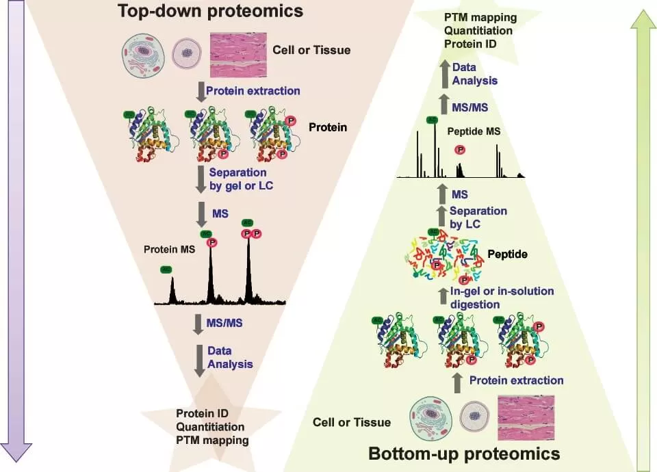
Figure 1. Conceptual comparison of top-down and bottom-up proteomics workflows.
Top-Down Proteomics: Analyzing Intact Proteins
Top-Down Proteomics refers to the method of mass spectrometry analysis conducted directly on intact proteins without any enzymatic digestion or chemical treatment. Unlike traditional methods that fragment proteins into peptides, Top-Down Proteomics directly examines whole proteins, enabling comprehensive characterization of their proteoforms—distinct protein variants arising from genetic variations, alternative splicing, and post-translational modifications (PTMs). This global analysis is performed using high-resolution mass spectrometry techniques, such as Fourier-transform ion cyclotron resonance (FT-ICR) or Orbitrap, which fragment intact proteins to reveal their molecular details.
The primary advantages of Top-Down Proteomics include the ability to avoid false-positive identifications and the simultaneous detection of multiple post-translational modification sites. Additionally, it effectively quantifies and distinguishes different proteoforms, capturing the true information of proteins. However, the approach faces significant challenges due to the proteome complexity in biological samples. The presence of high-abundance proteins, like albumin, can obscure low-abundance proteoforms, complicating separation techniques like liquid chromatography. Additionally, the large size of intact proteins demands advanced ion fragmentation methods and sophisticated bioinformatics for accurate data interpretation.
Workflow of Top-Down Proteomics
Conducting Top-Down Proteomics involves several critical steps, from sample preparation to data analysis. Below is a detailed guide to the entire process:
1. Sample Preparation
- Source Selection: Choose an appropriate biological sample, such as tissue, cell lysates, or body fluids. Ensure the sample is suitable for proteomic analysis.
- Protein Extraction: Isolate proteins from the sample using techniques like homogenization or centrifugation, depending on the sample type. Use buffers that maintain protein stability and prevent degradation.
- Concentration and Purification: Concentrate the protein solution using techniques such as precipitation (e.g., ammonium sulfate) or ultrafiltration to remove small molecules and contaminants.
2. Direct Protein Analysis
- Introduce Proteins to Mass Spectrometer: Load the intact proteins directly into the mass spectrometer. Ensure that the sample is in a suitable solvent that can be ionized effectively, often using a method like electrospray ionization (ESI).
- Ionization: Generate charged ions from the intact proteins for analysis. ESI is commonly used for Top-Down Proteomics because it can produce multiply charged ions from larger proteins.
3. Mass Spectrometry Identification
1) Fragmentation Techniques:
- Electron Transfer Dissociation (ETD): This method allows for the fragmentation of ions while preserving labile post-translational modifications, providing structural information about the protein.
- Ultraviolet Photodissociation (UVPD): UVPD uses high-energy ultraviolet light to induce fragmentation, offering another way to analyze protein structure.
2) Acquisition of Mass Spectra: Collect the mass-to-charge (m/z) ratios and intensity data for the intact proteins and their fragments. The mass spectrometer will produce spectra that represent the protein and its fragments.
4. Data Analysis
- Spectrum Interpretation: Use specialized software to interpret the mass spectrometry data. Analyze the mass spectra to identify protein peaks and their corresponding fragments.
- Protein Identification: Match the observed m/z values to known protein sequences using databases such as UniProt or NCBI. The identification may include assessing post-translational modifications and isoforms.
- Quantitative Analysis: If needed, use methods like label-free quantification or stable isotope labeling to compare protein abundance across different samples or conditions.
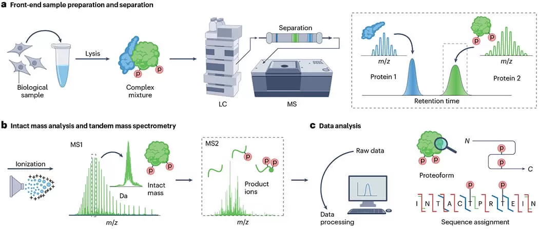
Figure 2. Representative top-down proteomics workflow from sample preparation to intact protein MS/MS and data analysis.
Characteristics of Top-Down Proteomics
- Preservation of Protein Integrity: Top-Down Proteomics allows for the simultaneous analysis of a protein's primary structure and post-translational modifications (PTMs). This capability provides a comprehensive view of the protein’s functional state.
- Isomer Resolution: This approach can directly differentiate between various proteoforms or isomers of a protein. By analyzing intact proteins, researchers can identify subtle differences that may significantly impact function.
- No Need for Database Searching: Proteins can be identified directly from mass spectrometry data, reducing the complexity associated with database searches. This advantage streamlines the identification process and minimizes potential errors that can arise during matching.
Limitations of Top-Down Proteomics:
- High Technical Requirements: The method demands high-resolution mass spectrometers and sophisticated data processing techniques. These requirements can pose challenges in terms of cost and accessibility, especially for laboratories with limited resources.
- Lower Analytical Throughput: Compared to Bottom-Up Proteomics, Top-Down methods generally have a lower throughput. This results in slower analysis times, making it more suitable for in-depth studies of a limited number of proteins rather than large-scale proteomic profiling.
Bottom-Up Proteomics: Fragmenting Proteins for Analysis
Bottom-up proteomics is the foundation of proteomics, of which shotgun proteomics is the most well-known. Bottom-up proteomics uses proteases such as trypsin to enzymatically digest proteins into smaller peptide fragments. These peptides are then analyzed by liquid chromatography-mass spectrometry (LC-MS), enabling high-throughput proteomic analysis of complex protein mixtures. The advantage of this approach is its scalability, making it ideal for large-scale proteome analysis in biomarker discovery and disease research. However, peptide-based analysis can lose post-translational modification (PTM) information, making the identification of protein variants more complicated.
Workflow of Bottom-Up Proteomics
Conducting Bottom-Up Proteomics involves a series of well-defined steps that encompass protein extraction, enzymatic digestion, peptide separation, and mass spectrometry analysis. Here is a comprehensive guide to the process:
1. Sample Preparation
- Sample Collection: Choose an appropriate biological sample, such as cell lysates, tissue homogenates, or body fluids. Ensure that the sample is suitable for proteomic analysis.
- Protein Extraction: Isolate proteins from the sample using methods like homogenization or centrifugation. Use a lysis buffer containing detergents and protease inhibitors to solubilize and stabilize the proteins.
- Protein Quantification: Determine the concentration of extracted proteins using methods such as the Bradford assay, BCA assay, or UV absorbance at 280 nm.
2. Enzymatic Digestion
- Select a Protease: Common proteases for digestion include trypsin, chymotrypsin, and LysC. Trypsin is the most widely used due to its specificity and efficiency.
- Digestion Process: Dilute the protein sample to reduce the concentration and facilitate digestion. Add the chosen protease in a specific enzyme-to-substrate ratio (typically 1:50 to 1:100) and incubate at an appropriate temperature (usually 37°C) for several hours or overnight. If necessary, consider performing a two-step digestion using different proteases for increased coverage of protein sequences.
3. Peptide Purification
- Stop Digestion: Once digestion is complete, stop the enzymatic reaction by adjusting the pH with formic acid or acetic acid.
- Peptide Cleanup: Remove salts, detergents, and other impurities using methods like:
- Solid-Phase Extraction (SPE): This method uses specialized columns to isolate and concentrate peptides.
- Ultrafiltration: Use membrane filters to concentrate and desalt the peptide solution.
4. Peptide Separation
Liquid Chromatography: Employ techniques such as reversed-phase liquid chromatography (RPLC) to separate peptides based on their hydrophobicity. This step reduces sample complexity and improves the quality of mass spectrometry data.
Use a gradient elution method to gradually increase the organic solvent concentration, facilitating the elution of peptides at different retention times.
5. Mass Spectrometry Analysis
- Ionization: Ionize the separated peptides using methods such as electrospray ionization (ESI) or matrix-assisted laser desorption/ionization (MALDI) to generate charged ions.
- Mass Spectrometry (MS): Introduce the ionized peptides into the mass spectrometer to measure their mass-to-charge (m/z) ratios. Collect the mass spectra for analysis.
- Tandem Mass Spectrometry (MS/MS): Select specific peptides for fragmentation to generate product ions. This provides additional information about the amino acid sequences and any post-translational modifications present.
6. Data Analysis
- Spectrum Interpretation: Use specialized software to analyze the mass spectrometry data. This includes identifying peptide sequences based on their m/z values and fragmentation patterns.
- Database Search: Match the identified peptide sequences to protein databases such as UniProt or NCBI to determine the corresponding proteins. Employ search algorithms that consider modifications and ensure accurate identification.
- Quantitative Analysis: Use label-free quantification methods or isotopic labeling techniques (such as TMT or iTRAQ) to quantify protein abundance across different samples or conditions.
Advantages of Bottom-Up Proteomics
- High Throughput and Scalability: Bottom-Up Proteomics is highly effective for analyzing large numbers of proteins in complex samples, making it ideal for high-throughput studies in various research fields.
- Established Protocols: There are well-established protocols and methodologies for Bottom-Up Proteomics, which facilitate the reproducibility and reliability of results.
- Enhanced Sensitivity: The method typically achieves higher sensitivity and precision, allowing for the detection of low-abundance proteins and a wide dynamic range of protein concentrations.
Limitations of Bottom-Up Proteomics
- Challenges in Analyzing Protein Modifications: Since proteins are digested into smaller peptide fragments, information about certain post-translational modifications (PTMs) may be lost during the digestion process. This limitation can hinder the ability to fully characterize the functional state of proteins.
- Limited Resolution of Protein Isoforms: Different isoforms of a protein may produce identical peptide fragments during digestion, making it challenging to distinguish between them. This limitation can lead to ambiguities in identifying specific proteoforms, which are crucial for understanding the diversity and function of proteins in biological systems.
Top-Down vs. Bottom-Up Proteomics: Which Strategy Should You Choose?
No single strategy is universally better. If the goal is proteoform-level characterization, intact mass measurement, or preservation of post-translational modification context on the same protein molecule, top-down proteomics is usually the better fit. If the goal is deep proteome coverage, large-cohort quantification, or routine analysis of complex biological samples, bottom-up proteomics is usually more practical. In many research settings, bottom-up is used for broad discovery, while top-down is used for proteoform-level follow-up and validation.
- Analysis Workflow: Bottom-Up Proteomics begins with the digestion of proteins into peptide fragments, while Top-Down Proteomics involves the direct analysis of intact proteins.
- Data Type: Bottom-Up Proteomics provides information at the peptide level, whereas Top-Down Proteomics yields comprehensive data about the entire protein.
- Application Areas: Bottom-Up methods are well-suited for large-scale proteomic screening, while Top-Down methods are ideal for structural and modification analyses of proteins.
- Technical Difficulty: Top-Down Proteomics requires more advanced technical capabilities, resulting in more complex data analysis.
Need help choosing the right proteomics workflow for your study? Explore our Proteomics Services including Quantitative Proteomics and PTM solutions to find a workflow that matches your sample type, cohort size, and research goals.
Conclusion
In summary, top-down and bottom-up proteomics answer different analytical questions rather than competing as direct substitutes. Top-down is stronger for intact proteoforms and PTM context, whereas bottom-up remains the preferred option for scalable protein identification and quantitative proteomics in complex samples.
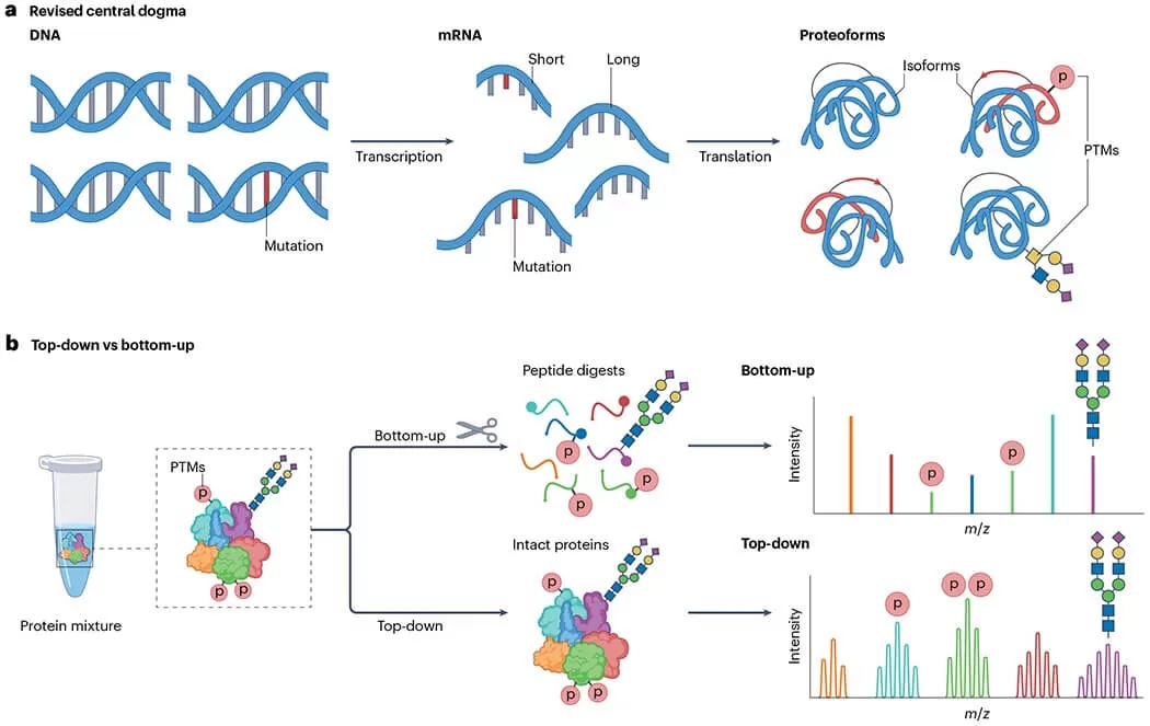
Figure 3. Proteoform generation and analytical differences between top-down and bottom-up proteomics.
FAQ
Q1. What is the main difference between top-down and bottom-up proteomics?
Top-down proteomics analyzes intact proteins directly, while bottom-up proteomics first digests proteins into peptides and then analyzes those peptides by mass spectrometry. Because of this difference, top-down preserves proteoform-level information, whereas bottom-up is better suited to large-scale peptide and protein identification.
Q2. When should I choose top-down proteomics?
Top-down proteomics is most useful when you need to characterize intact proteoforms, distinguish sequence variants or isoforms, or preserve the combinatorial context of post-translational modifications on the same protein molecule. It is especially valuable when structural detail matters more than throughput.
Q3. Why is bottom-up proteomics more commonly used?
Bottom-up proteomics is widely used because it is more scalable for complex samples, fits established LC-MS/MS workflows, and supports high-throughput protein identification and quantitative analysis. In routine proteomics projects, it is often the more practical strategy for broad proteome coverage.
Q4. Is top-down proteomics better for PTM analysis?
Top-down proteomics is often better when the goal is to preserve PTM context on intact proteoforms, because digestion in bottom-up workflows can break the linkage between modifications on the same protein molecule. However, bottom-up can still be highly effective for detecting and localizing many PTMs at the peptide level.
Q5. Can top-down and bottom-up proteomics be used together?
Yes. These two approaches are often complementary rather than mutually exclusive. Bottom-up proteomics can provide broad discovery and quantitative coverage, while top-down proteomics can be used for deeper characterization of selected proteins, proteoforms, or modification patterns.
Q6. Which approach is better for complex biological samples?
For highly complex biological samples, bottom-up proteomics is usually more practical because it offers higher throughput and broader coverage. Top-down proteomics can provide richer structural information, but it generally requires more advanced instrumentation, cleaner samples, and more specialized data analysis.
Read More in Proteomics
If you're comparing proteomics workflows, these related articles can help you go further—from PTM-focused strategy selection to quantitative workflows, data analysis, and foundational proteomics concepts.
For readers who want to move from workflow comparison into quantitative practice, this article explains how LFQ converts MS signals into protein abundance through feature detection, alignment, normalization, and peptide-to-protein quantification.
After understanding top-down and bottom-up workflows, many readers next need to know how proteomics data is actually processed. This guide connects raw files, identification, quantification, QC, statistics, and interpretation into one practical downstream workflow.
Since PTM context is one of the key reasons researchers care about top-down and related strategies, this article is a strong next read for users who want to understand PTM workflow design, site localization confidence, and false discovery control.
For readers who want to step back and strengthen their foundational understanding, this beginner-friendly article covers core proteomics concepts, protein basics, database use, and key quantitative ideas that support later workflow decisions.
This is a good next step for readers moving into more specific PTM workflows. It compares common phosphopeptide enrichment strategies and helps users connect high-level proteomics method choices with practical phosphoproteomics study design.
Take the Next Step in Your Proteomics Project
If you are moving from proteomics reading to project planning, our Proteomics Services can help you choose the right workflow for protein identification, quantitative profiling, PTM analysis, and downstream data interpretation based on your sample type and research goals.
Reference
- Gregorich, Z. R., Chang, Y. H., & Ge, Y. (2014). Proteomics in heart failure: top-down or bottom-up? Pflugers Archiv: European journal of physiology, 466(6), 1199–1209. https://doi.org/10.1007/s00424-014-1471-9
- Roberts, D. S., Loo, J. A., Tsybin, Y. O., Liu, X., Wu, S., Chamot-Rooke, J., Agar, J. N., Paša-Tolić, L., Smith, L. M., & Ge, Y. (2024). Top-down proteomics. Nature reviews. Methods primers, 4(1), 38. https://doi.org/10.1038/s43586-024-00318-2
Proteomics
Metabolomics
Multiomics
Contact Us
Name can't be empty
Email error!
Message can't be empty
CONTACT FOR DEMO
Next-Generation Omics Solutions:
Proteomics & Metabolomics
Have a project in mind? Tell us about your research, and our team will design a customized proteomics or metabolomics plan to support your goals.
Ready to get started? Submit your inquiry or contact us at support-global@metwarebio.com.
Ready to get started? Submit your inquiry or contact us at support-global@metwarebio.com.
Name can't be empty
Email error!
Message can't be empty
CONTACT FOR DEMO
Featured Multi-Omics Services
What's happening at Metware
Metabolite Extraction Methods for Metabolomics: Comparison, Selection, and Optimization
21
May
2026
Metabolite Extraction Methods for Metabolomics: Comparison, Selection, and Optimization
May 21, 2026
How Sample Pre-Treatments Affect Spatial Metabolomics: A Practical Guide to Perfusion, Fixation, FFPE, and Decalcification
May 14, 2026
Estrogen in Health and Disease: Types, Metabolism, Signaling, and Multi-Omics Research
May 13, 2026
